In this post we are going to deal with the various Ecg leads
we all know that the body acts as conductor of electricity & therefore recording electrode placed some distance from heart ,such as arms ,legs ,chest wall, are able to detect voltages conducted to these locations.
the usual way of recording ECG is by 12-lead ECG.
before going to depth lets first understand the difference b/w a ECG lead & an electrode.
- electrode-it simply means the metal plate used to detect electrical currents of heart in any location
- ECG lead-it shows the differences in voltages detected by electrodes.
12 LEAD ECG:
subdivided into 2 groups-
- limb leads-extremity leads-6 in number(in frontal plane)
- chest leads(precordial leads)-6 in number(in horizontal plane)
limb leads:has 2 groups-
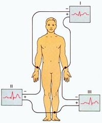
- bipolar limb leads:I, II, and III
- unipolar augmented leads : aVR,aVL & aVF
{note:bipolar leads were so named because they record difference in electrical voltage b/w 2 extremities
unipolar lead records electrical voltages at one location relative to an electrode with zero potential.}
bipolar limb leads:
- standard limb leads-I, II, and III
- recorded first
{note :here right leg electrode functions only as an electrical ground.although in theory it can be placed anywhere on the body. With a three-lead ECG, when one dipole is viewed, the remaining lead becomes the ground lead by default.}
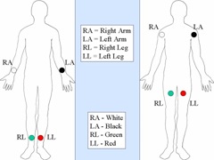
Lead I : records difference in voltages between the right arm and left arm electrodes, the left arm being positive.
Lead I =LA-RA
Lead II :records difference in voltages between the right arm and left leg electrodes, the left leg being positive.
Lead II =LL-RA
Lead III : records difference in voltages between the left arm and left leg electrodes, the left leg again being positive.
Lead III =LL-LA
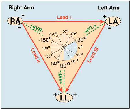
A diagrammatic representation of these three leads is termed Einthoven's triangle .
From the above three equations we get Einthoven's equation:
I +III = II.
It implies add the voltage in lead I to that in lead III & you get voltage in lead II.
{note: so it is a good practice to scan leads I, II, and III & if R wave in lead II does not seem to be sum of R wave in lead I & III , this may be clue that leads have not been mounted improperly}
augmented limb leads:
named as aVR,aVL & aVF :
- here 'a' stands for augmented
- V-voltage
- R ,L ,F-right arm ,left arm , & left foot respectively.
{note:
The augmented limb leads:
- The electrode positions for the "augmented limb leads" are exactly the same as the electrode positions for the "standard limb leads," above.
- An augmented limb lead measures electrical activity between the heart and the recording electrode.
- However, all of the electrodes used here are "positive" electrodes.
- Electrical activity traveling in a direction opposite to the lead being measured is determined as the average of the electrical activity measured at the other 2 electrodes. This sounds like a complex process, but it is all handled internally by the polygraph equipment doing the ECG recording.
- Mathematically, what is really happening is that the electrical activity being measured by any lead is "augmented" (amplified) by about 50% by this mathematical relationship.
- Using the acronyms VL (left shoulder), VR (right shoulder) and VF (left foot/leg):
aVL = VL - 0.5 (VR + VF) <---- simple formula for the "augmented left shoulder lead"
- The simple formula above for the "augmented left shoulder lead" (aVL) shows us that aVL is determined as the electrical activity recorded at the electrode on the left shoulder (VL) minus the average difference (-0.5) from the electrical activity measured at the other 2 electrodes (VR + VF).
- Now, let's see why this simple formula ends up amplifying the VL recording by 50%!!! In order to get started, we need to recognize that there exists a hypothetical "null" value at a central point over the heart where no fluctuations in electrical potential can be measured. In fact, this null point is demonstrated by algebraic summation of the electrical activity measured by all 3 limb leads... which adds to zero. So...
- VL + VR + VF = 0 (if we algebraically sum the electrical activity at all 3 electrodes, we get 0). Now, let's isolate the VL electrode...
- VL = - (VR + VF)..... Now, let's compare VL to the average value measured at the other 2 electrodes...
- 0.5 VL = -0.5 (VR + VF)... and what we see on the right side of this equation (-0.5 (VR + VF)) also appears on the right side of our simple aVL formula, above... so... let's replace the terms VR and VF in the simple aVL formula...
- aVL = VL - 0.5 (VR + VF) ..... becomes.... aVL = VL + 0.5 VL.... or, aVL = 1.5 VL... wow... so it appears, then, that aVL simply works out to be about 1.5 times the electrical activity recorded by VL. So, aVL is truly an augmented lead!
To summarize, then: An augmented limb lead is determined by measuring the electrical activity at a chosen limb lead, and then subtracting the average of the electrical activity measured at the other 2 limb leads. The net effect of this process is to augment the lead of interest by 50%.
The lead on the left arm is known as aVL (L for left), the lead on the right arm as aVR (R for right) and the lead on the left leg as aVF (F for foot). As the tracings are electrically "augmented" by comparison with the hypothetical null point over the center of the heart, at which no electrical potentials can be measured, the leads are referred to as "augmented leads."}
{note: now we know that
aVR +aVL +aVF=0
therefore it means -
sum of P waves in these three leads is 0
sum of QRS voltages is 0
sum of T wave voltages is 0.
{note :therefore ,when we scan the ECG for these leads & if the sum of waves in these leads does not equal 0 , then the leads might have been incorrectly placed.}
Hexaxial diagram:
the ECG leads have 2 important features:
- specific orientation(eg:lead I - oriented horizontally)
- specific polarity( eg" lead I - has positive polarity towards left side)
you can understand the remaining by looking at this diagram.
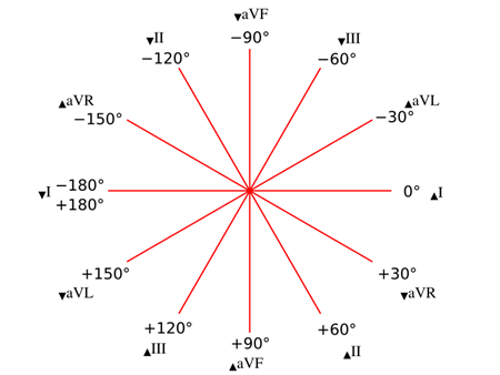
up arrow -positive pole & down arrow - negative pole.
It is used to help determine the heart's electrical axis in the frontal plane.{we will lran about this important concept in the next post}
{note: this is very important diagram & try to remember it BYHEART}
Chest leads:
- The precordial leads V1, V2, V3, V4, V5, and V6 are placed directly on the chest.
- Because of their close proximity to the heart, they do not require augmentation.
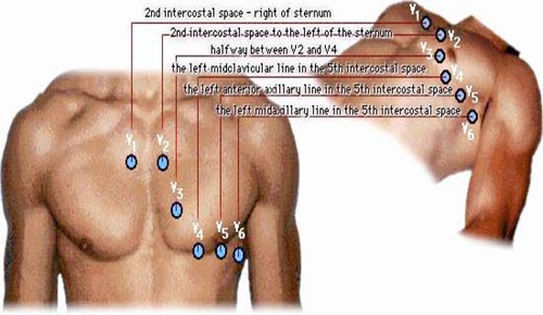
- The precordial leads view the heart's electrical activity in the so-called horizontal plane. The heart's electrical axis in the horizontal plane is referred to as the Z axis.
- Leads V1, V2, and V3 are referred to as the right precordial leads and V4, V5, and V6 are referred to as the left precordial leads.
- Wilson's central terminal is used for the negative electrode, and these leads are considered to be unipolar.
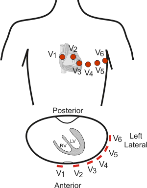
| Leads | group |
| V1, V2, V3, V4 | Anterior |
| I, AVL, V5, V6 | Left lateral |
| II, III, AVF | Inferior |
additional leads:
The "Lewis lead" :
- it is a bipolar lead with a negative electrode in the 2nd intercostal space on the right side and a positive electrode in the 4th intercostal space on the right side; a ground is placed in the 2nd intercostal space on the left side with all electrodes placed close to the sternum.
- used to detect atrial flutter waves when atrial flutter is suspected clinically but not definitely demonstrated on the standard 12 lead EKG.
- In order to create the Lewis Lead, move the right arm electrode to the right, second intercostal space adjacent to the sternum. Then move the left arm electrode to the right, fourth intercostal space adjacent to the sternum.
- The Lewis Lead is then read as Lead I on the EKG and, since in most patients it will be roughly perpendicular to the wave of atrial depolarization, flutter waves may be more apparent.
Internal leads: Electrode locations need not be in the "traditional" locations (ie. the arms, chest or back). Electrodes could also be located in the thoracic portion of the lumen of the esophagus, within the cardiac chambers, on the surface of the heart or within the AV bundle.
- The AV bundle electrogram gives very direct information about the major conducting elements of the heart.
We will learn the importance of these leads in diagnosing various disorders of heart in subsequent posts.
9 comments:
Very interesting. I would recheck one of the diagrams showing chest lead placement. I believe that V1 and V2 should be in the 4th Intercostal space and not the second as shown by the diagram. We were taught. Find the jugular notch, slid finger down to the Angle of Louis (2nd Intercostal Space), work down 2 ribs to the 4th Intercostal space. Either side of the sternam, Leads V1 and V2
MP
The description of the chest electrode positions is wrong. V1 and V2 should be in the 4th intercostal space, not the 2nd. V5 and V6 should be at the same horizontal level as V4, not in the 5th intercostal space.
Agree with the above coments. This picture of the V electrode placement should be taken down.
Dave Richley absolutely correct. Very poor post, only adding to confusion and poor practice of what is one of the most basic procedures. Shame to see it has not been corrected in five years!
Awful diagram of lead positions! Perfect to confuse unsuspecting students ...
It’s hard to find educated people on this subject, but you sound like you recognize what you’re speaking about! Thanks betfair online casino
How can I explain this to the world that there is a man who can cure HERPES, I was diagnosed for the past 1 year I have being into HERPES since 1 year, so I decide to look for help in the internet then I found a post write about this great man called DR. Yakubu , people say good thing about him that this man have cured, a lot of people in the internet, him has power's to cure HSV1 AND HSV2 I don’t believe that there is a cure for Herpes Simplex Virus because though there is no cure for herpes that what I have in mind HERPES had no cure well , my HERPES is negative through the using of herbal cured of DR yakubu , I contacted this man for help because of what i see in the internet. if you need his cured just email him now on dr.yakubuherbalhealingclinic@gmail.com , thank you DR. i will never stop shearing you testimony DR. Yakubu . him Can as well CURE THE FOLLOWING DISEASE:-1, HIV/AIDS, 2, Diseases of the hear 3, Malignant tumors 4. Cardiovascular diseases 5. Diabetes mellifluous 6.Influenza and pneumonia 7.Alzheimer's disease 8. hsv1 - hsv2. If you need LOVE SPELL. E.T.C contact him dr.yakubuherbalhealingclinic@gmail.com or you whatsapp him on +2348057353647...
Dr.yakubu 하나님은 내 삶을 회복시켜 주신 것에 대한 축복을 계속할 것입니다. 나는 사람들에게 증거를 계속 나눌 것입니다.
I started on COPD Herbal treatment from Ultimate Health Home, the treatment worked incredibly for my lungs condition. I used the herbal treatment for almost 4 months, it reversed my COPD. My severe shortness of breath, dry cough, chest tightness gradually disappeared. Reach Ultimate Health Home via their website www.ultimatelifeclinic.com . I can breath much better and It feels comfortable!
I was diagnosed with Bronchiectasis four years ago. For over two years, I relied on prescription medications and therapies, but unfortunately, the symptoms continued to worsen. My breathing became more laboured, and I experienced increasing fatigue and shortness of breath with even minimal activity. Last year, out of desperation and hope, I decided to try an herbal treatment program from NaturePath Herbal Clinic. Honestly, I was skeptical at first, but within a few months of starting the treatment, I began to notice real changes. My breathing became easier, the tightness in my chest eased, and I felt more energetic and capable in my daily life. Incredibly, I also regained much of my stamina and confidence. It’s been a life-changing experience I feel more like myself again, better than I’ve felt in years. If you or a loved one is struggling with Bronchiectasis, I truly recommend looking into their natural approach. You can visit their website at www.naturepathherbalclinic.com
info@naturepathherbalclinic.com
Post a Comment