Pages

Tuesday, July 22, 2008

Diseases of external ear (cont..)

In this post we will discuss about few miscellaneous conditions affecting external ear.

  • cerumen
  • keratosis obturans
  • external auditory canal cholesteatoma
  • atresia of meatus


Cerumen or impacted wax:

composed of-

  • sebaceous gland secretions
  • apocrine sweat gland secretions
  • desquamated epithelium & keratin
  • dirt

function:

  • protective as it lubricates the canal & due to its acidic nature

Motion of the ear canal provided by ordinary chewing movements together with the process of epithelial proliferation and lateral migration propel the cerumen outward in a self-cleansing manner.

complaints:

  • hearing loss or blocked sensation
  • tinnitus & giddiness due to impaction on tympanic membrane
  • Reflex cough:Arnold's cough reflex-due to irritation of auricular branch of vagus nerve.also known as Ear cough.

Diagnosis

  • Cerumen impaction is diagnosed by direct visualization with an otoscope.
  • seen as brown to blackish mass

wax

Treatment

Cerumen removal may be attempted by

  1. irrigation of the external auditory canal, with or without the use of ceruminolytics;
  2. by ceruminolytics alone;
  3. or by manual removal using a curette, forceps, or suction.

Keratosis obturans:

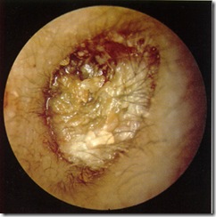
  • Keratosis obturans is characterized by a dense plug of keratin debris located primarily within the deep meatus.
  • with no associated erosion or necrosis of the surrounding bone.{THIS IS PRESENT IN external auditory canal cholesteatoma}
  • commonly seen b/w 5 & 20 years

etiology:

  • due to defect in epithelial migration
  • increased production of keratin debris
  • both

keratosis

Clinical features:generally BILATERAL & is PEARLY WHITE MASS.

  • Severe ear pain
  • Mild / moderate conductive hearing loss
  • Associated bronchitis / sinusitis - common{due to shared etiology of defective ciliary function resulting in defective migration}
  • Rarely otorrhea
  • Thickened tympanic membrane due to pressure of the keratin
  • Presence of granulations
  • Ballooning of the ear canal (bony reabsorption circumferentially widens the external bony canal)
  • On histological examination, the keratin plug displays a lamellar pattern(onion skin pattern) to the circumferential shedding of keratin squames from the auditory canal, with older layers being pushed centrally.

{note:

Keratosis obturans external auditory canal cholesteatoma
  • generally bilateral
  • unilateral
  • younger patients
  • older patients
  • origin is diffuse(along circumference of canal)
  • focal
  • generally otorrhea absent
  • present
  • also associated with bronchiectasis & chronic sinusitis
  • not associated
  • no osteonecrosis
  • presence of osteonecrosis & focal overlying epithelial loss present
  • managed successfully by regular aural-toilet
  • surgical intervention required

Treatment

  • Debridement of keratin debris (may require anesthesia)
  • Treatment of any underlying inflammation and/or infection
  • Periodical follow-up to remove reaccumulations

external auditory canal cholesteatoma:

we will deal with cholesteatoma in detail, in diseases of middle ear since more common in middle ear.

commonly involves post-inferior part of external auditory canal

etiology:

  • may be due to trapping of epithelium below level of skin during previous episode of otitis externa.
  • margins of it secrete enzymes & also due to pressure effects results in bone erosion
  • so often Facial nerve is exposed & paralysed.

iatrogenic external auditory canal cholesteatoma:

  • generally at anterior angle of tympanic membrane
  • etiology:due to incorrect repositioning of skin flaps at end of procedure

Acquired atresia & stenosis of meatus:

causes-

  1. infections:chronic otitis externa - important cause
  2. burns
  3. trauma

treatment:

meatoplasty {In a meatoplasty, an incision is made immediately behind the meatus, and a portion of the conchal cartilage is excised.}


Wednesday, July 9, 2008

Surgery-Abdomen introduction

First we will deal with various common abdominal disease presentations, & history taking along with the important clinical findings to be noted.

Abdominal disease presentations:

  • non-leading & misleading symptoms
  • minimal signs
  • require elaborate investigations

different complaints:

  • pain
  • dysfunction of diseased organs
  • lump
  • distension
  • general disturbance depending upon disease process

Abdominal pain:pain may be produced by-

  • somatic structures (abdominal wall ,parietal peritoneum)-these are innervated by somatic nerves & they are under cortical control.therefore pain is well localised & precise.
  • visceral structures(gut, solid alimentary organs, genitourinary organs)-these are under autonomic control(not under control of cerebral cortex) & hence pain is diffuse

{note:autonomic nerves go to hypothalamus where pain cannot be sensed ,so now you might get a doubt then how visceral pain is felt ?

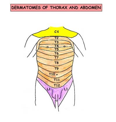
    • Actually the somatic & autonomic nerves both travel closely in a spinal nerve & this leads to some amount of leakage of current from autonomic to somatic nerve ,so now the actual visceral pain is felt by brain as if coming from the area supplied by that particular somatic nerve ,this is referred(radiating) pain.i.e, it is felt on same dermatomal segment of abdominal wall.
    • visceral pain felt only when organ is stretched ,spastic or ischemic or due to stretch of mesentry
    • pain can be modified by neighboring somatic structures:

eg-in appendicitis -first pain in umbilical region ,then depending upon degree of inflammation exudate leaks out & collects in right illiac fossa peritoneum (which is supplied by somatic nerves) &hence causes pain in right illiac fossa}

  • viscera in somatic compartment(pancreas, kidney etc)-to explain about how these organs feel pain let me tell you with an example-pancreas is supplied by Sympathetic nerves T8,T9,T10.so as you see the given illustration referred pain will be above umbilicus when pancreas is inflamed.as time passes the inflammatory exudate leaks out into somatic compartment(i.e, retroperitoneum) & irritates the somatic nerves & causes back pain also.

abd dermatomes

DIVISION ARTERY

VEIN

LYMPHATICS

SYMPATHETIC

PARASYMPATHETIC

FOREGUT:

  • Oesophagus
  • Stomach
  • Proximal half of duodenum (up to common bile duct (CBD))
  • Liver
  • Pancreas
CELIAC ARETERY

PORTAL VEIN

Spleenic vein

Gastric vein

CELIAC NODES CELIAC GANGLIA
(T6 to T8)
VAGUS

MIDGUT:

  • Distal half of duodenum (from CBD)
  • Jejunum
  • Ileum
  • Appendix
  • Caecum
  • Ascending colon
  • Right 3/4 of transverse colon
SUPERIOR MESENTERIC ARTERY SUPERIOR MESENTERIC VEIN SUPERIOR MESENTERIC NODES SUPERIOR MESENTERIC GANGLIA
(T9 to T10)
VAGUS

HINDGUT:

  • Left 1/4 of transverse colon
  • Descending colon
  • All of rectum down to ano-rectal line
INFERIOR MESENTERIC ARTERY INFERIOR MESENTERIC VEIN INFERIOR MESENTERIC NODES

GANGLIA

HYPOGASTRIC PLEXUS

(T11 to T12 ,L1)

PELVIC SPLANCHNIC NERVES

[note:

so pain in gut viscera is referred to their respective dermatomes ,especially at midline & only in severe cases to sideways

  1. pain in foregut-above umbilicus
  2. midgut-at umbilicus
  3. hindgut-below umbilicus]


Gall bladder pain:

pain along T7 -T9 segments(right hypochondrium to inferior angle of right scapula)

Diaphragm pain:

along C3,4,5 (pain over shoulder)

Urinary organs pain:

along dermatomes L1 & L2(along genitofemoral nerve)

retroperitoneal visceral pain:

pain is felt in back in addition to referred pain on sensory dermatomes

eg-pain in pancreas.

Nature of pain:varies with nature of disease

  • continous pain -inflammatory
  • colic (intermittent ,gripy ,in wave form) -spastic conditions.

Dysfunction:

In gut disease it results in Dyspepsia -it is a very vague term literally meaning -difficulty in digestion.

patient may complain of -

  • lack of appetite
  • nausea
  • vomiting
  • diarrhea
  • constipation ,etc

now let us consider each abdominal organ dysfunction separately.

stomach:

  1. mild dysfunction results in -anorexia
  2. moderate - nausea
  3. severe - vomiting

note:stomach has property of receptive relaxation(to receive food from oesophagus) & propulsive expulsion(to expel into duodenum).

  • bloating sensation(due to absence of receptive relaxation)-patient feels heavy even with small amount of food ,since there is no relaxation of stomach.

small bowel:

  • malabsorption,diarrhea

colon:

  • altered bowel habits
  • discharge of mucus per rectum

liver:jaundice

urinary organs:disturbance in micturation

female reproductive organs:menstural disturbance.

Modified presentation

due to exaggerated sympathetic response to some organs, especially stomach.

let me explain it to you with an example:

  • suppose small bowel is diseased ,so to recover it needs rest from its usual work. so for this to occur the stomach causes symptoms like anorexia , nausea ,vomiting .so as a result the person wont like to eat anything & thus his small bowel gets some rest & it recovers fast.
  • suppose the stomach doesn't make these manifestations to occur ,then the person keeps on eating & there is no rest for small bowel & its condition detiorates.
  • Hence stomach acts as a BIG BROTHER of abdomen.
  • sometimes its response is greater than the diseased organ & disease is mistaken as gastric disorder. so you should keep in mind about the other possibilities of each presentation.

presentation as lump:

site of lump indicates site of its origin unlike pain.

now lets go to clinical examination:

  • detailed history taking
  • general examination
  • abdominal examination
  • examination of scrotum ,supraclavicular nodes & spine.

detailed history taking:

points to be enquired

pain:

  • site( initial & present)
  • nature
  • aggravating & relieving factors
  • relation to food ,vomiting ,defecation
  • periodicity

presenting features of dysfunction of viscera

  • anorexia ,nausea ,vomiting(stomach)
  • diarrhea(small bowel)
  • altered bowel habits .mucus/ blood in stools (colon)
  • frequency ,difficulty in micturation ,haematuria (urinary organs)
  • menstural disturbance (female genital tract)
  • jaundice(liver)
  • any general disturbance like fever ,loss of weight (T.B. ,malignancy ,etc)

lump:

  • initial site of appearance
  • rate of growth
  • associated with pain or not
  • preceding history of abdomen disease.

general examination.

PICCKLE ,heart & lung sounds.

P-pallor(check lower palpebral conjunctiva)

I-icterus/jaundice(upper bulbar conjunctiva)

C-cyanosis(tongue)

C-clubbing

K-koilonychia

L-lymphadenopathy

E-edema

abdominal examination

Inspection:

skin & subcutaneous tissue:

  • abdominal lump
  • superficial engorged veins:
features portal obstruction inferior vena cava obstruction superior vena cava obstruction
position situated around umbilicus (caput medusa) on sides of abdomen on sides of abdomen
direction of blood flow away from umbilicus from below upwards from above downwards

engorged veins

note:direction of blood flow is known by following technique-

  • two index fingers of both hands are placed close together on engorged vein below umbilicus
  • portion of vein is emptied by milking it with one of the index fingers
  • remove the upper finger, the vein remains collapsed if flow is from below upwards .vein fills quickly if flow is from above downwards.
  • repeat the same process of emptying the vein , but now remove the lower finger.the vein remains collapsed if flow is from above downwards .vein fills quickly if flow is from below upwards.

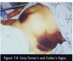
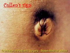
Cullen's sign & Grey-Turner's sign:

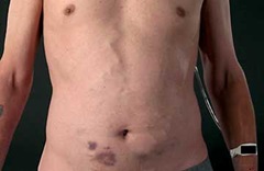
  • Cullen's sign is ecchymosis (puplish blue discolouration) of the skin around the umbilicus.
  • Grey-Turner's sign is ecchymosis in flanks.
  • mainly associated with hemorrhagic pancreatitis (in 1 to 2 % cases seen)
  • (typically after 2 to 3 days after acute pancreatitis)
  • represent retroperitoneal hemorrhage that has dissected through fascial planes to skin

cullens sign Cullen

also seen in:

  1. ruptured ectopic pregnancy ,severe trauma
  2. leaking or ruptured abdominal aortic aneurysm
  3. any condition associated with bleeding into retroperitoneal space

It is caused by pancreatic enzymes that have tracked along the falciform ligament and digested subcutaneous tissues around the umbilicus.

scar:

  • linear scar -healing by primary intention
  • irregular scar -indicating wound infection
  • keloid , hypertrophic scar.

hard subcutaneous nodules near umbilicus:Sister Mary Joseph Nodule

Sister Mary Joseph Nodule

  • She was first to notice that a 'nodule' in the umbilicus was often associated with advanced malignancy
  • Presents as firm, red, non-tender nodule
  • Results from spread of tumour within the falciform ligament
  • 90% of tumours are adenocarcinomas
  • Commonest primaries are stomach and ovary
  • Primary tumour is almost invariably inoperable

abdomen distention:5F's

  1. fetus
  2. faeces
  3. flatus(gas)
  4. fluid
  5. fat

note : among these only in fat distention ,umbilicus is deeply inverted & in others it is flat or everted.(reason:umbilicus is a scar ,which is bound to linea alba & when fat increases, it fills around the umbilicus as subcutaneous fat & thus causes raising of level of skin around it)

umbilicus:

tanyol's sign:umbilicus is displaced upwards by swelling arising from pelvis or downwards by ascites.

any swelling on one side of abdomen will push umbilicus to opposite side.

movements:

respiratory - localised limitation indicates subjacent inflammation

peristaltic -

observed in pyloric stenosis obstruction in small bowel obstruction in large bowel
features rounded prominence will be seen travelling slowly from left costal margin to right ladder pattern opposite movement to that of pyloric stenosis

pulsatile:

  • aortic aneurysm - expansile pulsation
  • tumor in front of abdominal aorta - transmitted pulsation

palpation:

tenderness(pain on touch):

disease tender spot location
gastric ulcer mid-epigastrium below xiphoid process
duodenal ulcer 1&1/2 inch to right of midline on transpyloric plane
cholecystitis
  • Palpation under the right costal margin whilst asking the patient to breathe in causes pain as the inflammed gallbaldder touches with the palpating hand.(Murphy's sign)
  • cartilage of right 8th rib becomes tender
  • hyperaesthesia below the right scapula, called Boas' sign.
appendicitis McBurney's point(McBurney's point refers to the location of the base of the appendix. It lies one third of the way along an imaginary line drawn from the anterior superior iliac spine to the umbilicus.)-this is McBurney's sign.

rigidity & guarding(stiffness):in both there is reflex contraction of abdominal wall

  • in rigidity ,it is present at rest &
  • in guarding it appears due to provocation from pressure of examining fingers

lump:

  • character(consistency)
  • its anatomical plane(intra-abdominal or parietal) :

abdominal muscles are made taut by asking patient :

  1. to raise his shoulders from bed with arms folded on his chest 'the rising test'
  2. to raise both extended legs from bed -'leg lifting test'(carnett's test)
  3. try to blow out with nose & mouth shut.

results:if swelling is-

  • parietal -swelling will become more prominent & will be freely moveable over the taut muscle.
  • parietal but fixed to abdominal muscles = swelling will not be moveable over the taut muscle.(eg: recurrent fibroid of paget & haematoma in rectus muscle)
  • intra-abdominal-disappears or becomes smaller.

note:if swelling moves vertically with respiration it is intra-abdominal.

Palpation of abdominal organs:

  • stomach
  • liver
  • spleen
  • gallbladder
  • kidney
  • pancreas
  • colon

stomach:

  • normally cannot be palpated.
  • in cases of pyloric stenosis-look for
    1. visible peristalsis
    2. succussion splash
    • the stomach becomes distended with fluid (ingested liquid ,saliva & gastric fluid) when the pylorus is obstructed by tumor or cicatricial stenosis.
    • when the diaphragm of stethoscope is placed medial to left costal margin & patient is shaken from side to side by the lower ribcage ,a splashing sound is heard ,that is pathognomic for this condition. books_0051
    • the test must be carried out after a 4hr fast.(since Succussion splash may be heard in normal subjects for up to 3 hours after a meal).
    • A positive succussion splash indicates a hollow area containing both fluid and gas.
    • For abdominal examination, the examiner's hand is placed in the area to be examined, a soft pressure is applied, and then the examiner gives short ,sudden ,jerky movements with fingers. This will, if the sign is positive, result in a palpable or audible splash.
    • For thoracic examination, auscultates while gently moving the patient side to side on the examining table.
    • A thoracic succussion splash is found in a pneumohydrothorax (air and fluid in the chest cavity) or rarely in hemopneumothorax.

lump in pyloric region:

  • congenital pyloric stenosis - thick pylorus
  • carcinoma - mass is irregular , hard with varying degrees of mobility
  • leaking perforation of peptic ulcer - firm ,less mobile ,more tender

{absence of lump by no means excludes carcinoma of stomach}

Liver:

  • children upto end of 3 years - palpable
  • adults - not palpable

Gall-Bladder:

when distended felt as tense globular swelling projecting downwards & forwards from below liver just lateral to outer border of right rectus muscle.

Courvoisier's law

  • It states that, in the presence of jaundice, an enlarged gallbladder is unlikely to be due to gallstones; rather carcinoma of the pancreas or the lower biliary tree is more likely.
    • reason:
  • Gallbladder distension has been thought to occur because the distal malignant obstruction leads to chronically elevated intraductal pressures. This contrasts with obstructions caused by stones, which are associated with chronic cholecystitis and fibrosis of the gallbladder wall, which precludes distension.
  • In addition, stones may cause only partial obstructions (related to a "ball-valve" action of the stone) leading to less consistent intraductal pressure elevations, and less gallbladder dilatation.

Exceptions to Courvoisier's Law

1. Oriental cholangiohepatitis
2. Double stone impaction (one in cystic duct, other in Common Bile Duct)
3. a pancreatic calculus obstructing ampulla of vater
4. Mucocele of gall bladder due to an impacted stone at neck of gall bladder.

Spleen:

  • spleen must be atleast two times larger than normal size to be detected clinically.
  • splenic swelling:sharp anterior border where 1 or 2 notches felt (characteristic of splenic swelling)

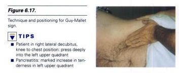
Pancreas:best way to palpate it:

  1. turn patient to right.
  2. hips & knees flexed
  3. left subcostal & epigastric regions deeply palpated.

if tenderness present - indicates pancreatitis (this is Guy -Mallet sign)

guy mallet sign

this pain & tenderness often radiates to left shoulder - Kehr's sign

Renal:

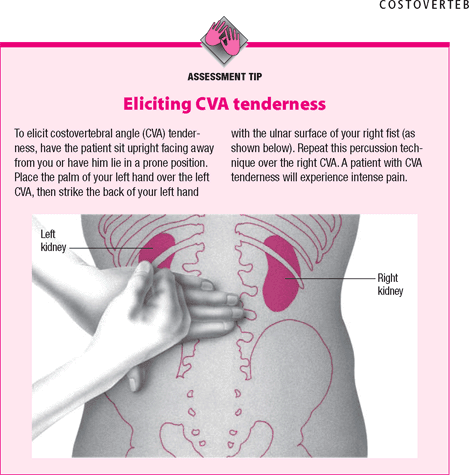
renal angle :check this area for signs of renal disease

  • An area located on either side of the human back between the lateral borders of the erecter spinae muscles and inferior borders of the twelfth rib, so called because the kidney can be felt at this location.
  • check for any dullness ,tenderness ,fullness.

cva

percussion:

evaluate-

  • free fluid in abdomen (by shifting dullness)
  • presence of gas filled viscus in front of any lump: eg-if anterior to stomach -dull note on percussion.
  • if posterior to stomach-resonant note on percussion
  • borders of solid organs mapped out by their dull note

ascites ovarian cyst
resonance anteriorly flanks
dullness flanks anteriorly
shifting dullness present absent

Spleen: to evaluate splenomegaly -

Castell's sign -

  • percussion in the lowest intercostal space(8th or 9th) in the left anterior axillary line.
  • In normals this area is resonant( due to the air-filled stomach or splenic flexure of the colon.)in inspiration and expiration.
  • With splenic enlargement this area becomes dull on maximal inspiration.

percussion over traube's space:

  • It's a crescent-shaped space, encompassed by the lower edge of the left lung, the anterior border of the spleen, the left costal margin and the inferior margin of the left lobe of the liver.
  • Thus, its surface markings are respectively the left sixth rib, the left anterior axillary line, and the left costal margin.

traube space

  • Underneath Traube's space lies the stomach, which produces a tympanic sound on percussion.
  • If percussion over Traube's space produces a dull tone, this might indicate splenomegaly

note:Castell’s has been shown to be superior in sensitivity to other spleen percussion signs as well as palpation, which is not likely useful due to the extreme enlargement necessary to feel the spleen below the costal margin.

auscultation:

• Systolic bruit heard over an artery indicates stenosis of the underlying artery. Systolic bruit may be heard also over very vascular intra-abdominal tumors.

• Venous hum is rarely heard. When present, it is a sign of venous collaterals developed secondary to portal hypertension.

Kenawy's sign:

  • seen in splenomegaly due to portal hypertension
  • hear a venous hum louder on inspiration with stethoscope placed below xiphoid process
  • reason:due to spleen being compressed during inspiration results in engorgement of splenic vein & hum is heard

Left supraclavicular lymph nodes:

enlargement of these group of nodes occurs in carcinoma of stomach - Troisier's sign.

This is end of todays post ,to sum up I will write the important high yield facts here:

    • Cullen's sign is ecchymosis (puplish blue discolouration) of the skin around the umbilicus.Seen in hemorrhagic pancreatitis.
    • Grey-Turner's sign is ecchymosis in flanks. seen in hemorrhagic pancreatitis.
    • Guy -Mallet sign : tenderness present in left subcostal & epigastric regions when deeply palpated.seen in pancreatitis.
    • Kehr's sign :pain & tenderness often radiates to left shoulder in pancreatitis.

  • Sister Mary Joseph Nodule : a 'nodule' in the umbilicus seen especially in advanced stomach cancer.

    • tanyol's sign: umbilicus is displaced upwards by swelling arising from pelvis or downwards by ascites.

  • Boas' sign :hyperaesthesia below the right scapula in cholecystitis
  • Murphy's sign :Palpation under the right costal margin whilst asking the patient to breathe in causes pain as the inflammed gallbaldder connects with the palpating hand

    • Castell's sign - percussion in the lowest intercostal space(8th or 9th) in the left anterior axillary line. causes a dull note in splenomegaly
    • Kenawy's sign :venous hum louder on inspiration with stethoscope placed below xiphoid process seen in splenomegaly due to portal hypertension

Sunday, July 6, 2008

Rosettes & Pseudorosettes - Neuropathology

This is one of the important topics in neuropathology  for diagnosis of brain tumors.To get the basic concept about different type of rosettes & in which tumors they occur & understand the topic better ,I have a nice article published in American Journal of NeuroRadiology.

You can download the pdf file from 4shared.

Friday, July 4, 2008

Inflammation of Ear canal

Based on aetiology:

infective type:

  • Bacterial
  1. localised otitis externa(furuncle)
  2. diffuse otitis externa
  3. malignant otitis externa
  • Fungal
  1. otomycisis(fungal otitis externa)
  • viral
  1. herpes zoster oticus
  2. otitis externa haemorrhagica


reactive type:

  • eczematous otitis externa
  • seborrhoeic otitis externa
  • neurodermatitis
Continue to read, by clicking on READ MORE.

furuncle:

Aetiology-staphylococcal infection of hair follicle

clinical features-

  • severe pain & tenderness which are out of proportion to size of furuncle
  • movements of pinna painful
  • jaw movements painful
  • if occludes canal ,conductive deafness

furuncle

Examination-

  • primary lesion is often a small pustule that may enlarge to become a furuncle
  • edema, erythema, tenderness and
  • fluctuance if abscess forms
  • periauricular lymph nodes enlarged & tender

treatment-

  • early stages(without abscess)-
  1. systemic antibiotics,
  2. analgesics,
  3. local heat,
  4. ear pack of 10% ichthammol glycerine (reduces edema)
  • late stage(with abscess)- incision &drainage and same as above

Extension of the infection to the pinna and periauricular soft tissues may warrant parenteral antibiotic therapy.

{note:

since as you know that hair follicle present only in outer third of ear canal(cartilagenous part) furuncle occurs in outer part only

recurrent furunculosis - suspect diabetes as cause}

Diffuse otitis externa:

  • can involve whole of canal ,tympanic membrane & pinna
  • also known as ’swimmers ear’

cause

  • The most common precipitant of otitis externa is excessive moisture. Excessive moisture removes cerumen and increases the pH of the external auditory canal, situations that provide a good setting for bacterial growth. and
  • trauma (due to scratching ,vigorous cleaning unskilled instrumentation)
  • both of the above two impair the canal's natural defenses,& allow bacterial entry into ear canal
  • The most common causative organism is Pseudomonas species and Staphylococcus aureus. Other pathogens less commonly cultured include Proteus mirabilis, Streptococci species, coagulase negative Staphylococci, and various gram negative bacilli.

clinical features-

Acute otitis externa- Chief symptoms include Otolagia& Otorrhea.

can further be divided into

  • preinflammatory:

The preinflammatory stage begins with itching, edema and a full sensation in the ear.

  • acute inflammatory stages. It is again divided into mild, moderate, or severe.
  1. As the infection progresses increased itching and pain ensues with mild erythema and edema on physical exam, however the canal lumen remains patent with cloudy secretions.
  2. During the moderate phase, the itching and pain intensify, and although the lumen remains patent, significant edema and debris decrease its size. Secretions are noted to be exudative and more profuse.
  3. Finally, in the severe stage of the disease, the pain is usually intolerable and is often intensified by manipulation of the skin and soft tissue around the ear. The lumen of the EAC may be obliterated by edema, debris and purulent otorrhea.
  • The auricle and periauricular soft tissues are often involved, and regional lymph nodes may become palpable.Fever may be present, but if it exceeds 38.3°C (101.0°F), more than simple local otitis externa should be considered. Lymphadenopathy just anterior to the tragus is common.
  • In patients where the disease does not resolve after treatment, a subacute or chronic form may occur.

treatment:

  • ear cleaning is very important
  • analgesics like codeine or NSAIDs used
  • topical antibiotics
  1. An aminoglycoside combined with a second antibiotic and a topical steroid such as neomycin, polymyxin B, and hydrocortisone used to be the most commonly prescribed topical antibiotic. However, caution must be used to watch for a hypersensitivity reaction to the neomycin and ototoxicity from the aminoglycoside. This preparation should not be used in cases of a perforated tympanic membrane, which is not always easy to determine in cases of otitis externa.
  2. Fluoroquinolones are not assoiciated with ototoxicity and ofloxacin is safe in cases of a perforated tympanic membrane.
  3. Ofloxacin otic is the drug of choice when a perforated tympanic membrane cannot be ruled out.
  • Systemic Treatment.: Oral antibiotics are rarely needed but should be used when otitis externa is persistent, when associated otitis media may be present or when local or systemic spread has occurred. The latter should be suspected if the patient's temperature is higher than 38.3°C (101.0°F), if initial pain is severe or if regional lymphadenopathy of the preauricular or anterior or posterior cervical chains is present.

{note:Currently, the only ototopical drop approved by the FDA for use in an open middle ear is ofloxacin. }

Chronic Otitis externa: Is defined when the duration of the infection exceeds 4 weeks or when more than 4 episodes occur in 1 year.

clinical features-

  • characterized by irritation & strong desire to itch.
  • discharge is scanty &dry up to form crusts
  • meatal skin thick & swollen
  • rarely skin becomes hypertrophic leading to meatal stenosis (CHRONIC STENOTIC OTITIS EXTERNA)

{note: discharge in otitis externa is serous type & in otitis media is mucoid type (this is due to presence of mucous membrane in middle ear)

treatment:

  • reduce swelling=10% ichthammol glycerine
  • reduce itching=antibiotic & steroid cream
  • chronic stenotic otitis externa=surgical procedures to enlarge and resurface the EAC, such as conchal meatoplasty, are indicated.

Malignant otitis externa:"Osteitis of the base of the skull,"

Necrotizing or malignant otitis externa is a life-threatening extension of external otitis into the mastoid or temporal bone.

Known as Malignant due to high risk of Complications.

Aetiology-

  • Most commonly caused by Pseudomonas aeruginosa, (98%)
  • occurs most often in elderly patients with diabetes mellitus. However, all immunocompromised patients, especially those with human immunodeficiency virus (HIV) infection, are at risk.
  • begins as an acute otitis externa and frequently progresses to a skull base osteomyelitis with resultant cranial neuropathies.

Clinical features-

  • The typical patient is an elderly diabetic with poor metabolic control and evidence of otitis externa not responding to the usual local therapy.
  • The typical complaints are deep-seated aural pain, discharge, and fullness.
  • A history of diabetes or an immunocompromised state (neoplasm, immunosuppressive therapy, HIV, etc.) should be elicited.
  • Examination of the involved ear canal reveals inflammation and granulation tissue at the bony cartilaginous junction. Purulent secretions are common, and excessive inflammation may occlude the canal and obscure the TM

{note:One of the hallmarks of necrotizing external otitis is granulation tissue in the external auditory canal, especially at the bone-cartilage junction.}This otoscopic finding is of extreme importance.

  • The infection may extend to the cartilaginous skeleton of the ear canal and through Santorini's fissures to reach the temporal bone, causing osteitis. One of the hallmarks of this extension is granulation tissue in the bone-cartilage junction of the external auditory canal.
  • Cranial nerve involvement may appear as early as one week after the onset of symptoms, with the facial nerve most commonly involved, followed by X and XI.

Diagnosis

Special alertness is required when external otitis is refractory to treatment and patients complain of severe otalgia, especially at night. The diagnosis of necrotizing external otitis is based on the clinical presentation and confirmed by laboratory tests and imaging studies.

LABORATORY TESTS

Mandatory laboratory tests include an erythrocyte sedimentation rate (ESR), white and red blood cell counts, glucose and creatinine levels, and culture of ear secretions. The ESR is typically elevated in necrotizing external otitis; therefore, it is a useful indicator of treatment response. Before topical or systemic antibiotic therapy is started, ear secretions should be cultured, because susceptibility patterns may change after the initiation of treatment (i.e., bacteria might become resistant to an antibiotic during treatment).Pathologic examination of granulation tissue removed from the external auditory canal is essential to exclude malignant processes, which may present as nonresponding inflammatory disease.

IMAGING STUDIES

Imaging to prove the extension of infection to bony structures is generally necessary to establish the diagnosis of necrotizing external otitis.

Imaging modalities include

  1. computed tomographic (CT) scanning,
  2. technetium Tc 99m medronate methylene diphosphonate bone scanning, and
  3. gallium citrate Ga(gallium) 67 scintigraphy.

CT scanning is used to determine the location and extent of diseased tissue.

  • The temporal bone is the first bone to be affected, with imminent involvement of the petrous apex and mastoid.
  • Extratemporal bone extension has become rare since the introduction of powerful antibiotics.
  • In evaluating the CT scan, it is important to remember that at least one third of bone mineral must be lost before radiologic changes become apparent; conversely, bone remineralization continues long after the infection is cured.
  • Thus, as related to the infectious process, pathology is late to appear on the CT scan and late to disappear.
  • These factors limit the usefulness of CT scanning as a follow-up tool.

Tc(technitium) 99m bone scanning:

  • Both osteoclasts and osteoblasts absorb 99mTc.
  • Hence, bone scanning can locate a pathologic process in bone but is not informative about the nature of the process (infectious or other).
  • Because the 99mTc scan remains positive as long as bone repair continues, this imaging modality is not helpful in follow-up.

gallium citrate Ga(gallium) 67 scintigraphy. :

  • Since 67Ga is absorbed by macrophages and cells of the reticuloendothelial system, scanning with this radioisotope is a sensitive measure of ongoing infectious process .
  • If 67Ga scintigraphy is available, it should be used for initial diagnosis and as a follow-up tool.
  • By using imaging modalities in combination, it is possible to prove that the temporal bone is afflicted (CT scanning and 99mTc bone scanning) with an infectious process (67Ga scintigraphy).

Treatment:

  • The excellent antipseudomonal activity of fluroquolones has generally made them the treatment of choice for necrotizing otitis externa,
  • although a combination of a beta-lactam antibiotic and aminoglycoside is also effective.
  • In severe cases, a prolonged course of parenteral antibiotics may be needed, but the excellent gastrointestinal absorption of the fluoroquinolones allows milder infections to be treated with a two-week course of oral therapy.
  • Treatment should also include surgical debridement of any granulation or osteitic bone

otomycosis:

cause-

  1. aspegillus niger ,
  2. aspergillus fumigatus ,or
  3. candida albicans.

The most common pathogen is Aspergillus (80 to 90 percent of cases), followed by Candida.

Classically, fungal infection is the result of prolonged treatment of bacterial otitis externa that alters the flora of the ear canal. Mixed bacterial and fungal infections are thus common

Clinical features :

  • the most common symptoms of otomycosis are pruritus deep within ear& an irresistible urge to scratch.
  • The itching generally progresses to dull pain with or without drainage.
  • The accumulation of fungal debris in the inflamed, narrowed canal often leads to a complaint of hearing loss.
  • Tinnitus is also a common presenting complaint.
  • however, with severe infections pain will also occur

examination with otoscope:Physical examination generally demonstrates canal erythema, mild edema and the presence of white, gray, or black fungal debris within the canal.

fungal mass is seen as "wet piece of filter paper"

  • A.niger-black headed filamentous growth.

otomycosis

  • A.fumigatus-pale blue or green
  • Candida-white or creamy deposit.

otomycosis2

treatment:

  • Cleansing of the ear canal by suctioning is a principal treatment.
  • Acidifying drops, given three or four times daily for five to seven days, are usually adequate to complete treatment.
  • Because the infection can persist asymptomatically, the patient should be reevaluated at the end of the course of treatment. At this time any further cleansing can be performed as needed. If the infection is not resolving, over-the-counter clotrimazole 1 percent solution (Lotrimin), which also has some antibacterial activity, can be used
  • Nystatin is effective against candida

Herpes Zoster Oticus (Ramsay Hunt syndrome) -some information taken from drtbalu wonderful educational site on ENT.

Ramsay Hunt syndrome is a disease affecting the external auditory canal associated with the following symptom complexes:

1. Lower motor neuron type of facial nerve palsy

2. Herpetic blisters of the skin of the external auditory canal

3. Otalgia

Pathophysiology:

The primary pathophysiology is located in the geniculate ganglion of the facial nerve. Geniculate ganglion is found to be affected by Human Herpes virus type 3 i.e. (Varicella zoster virus). Varicella zoster virus have been identified from tears of these patients by polymerase chain reaction. Infact Varicella zoster virus have also been identified from tears of patients with Bell's palsy.

Clinical features:

The classic Ramsay Hunt syndrome is associated with

1. Patient has deep seated pain in the affected ear. The pain is intermittent in nature, radiating towards the pinna of the ear. There is associated diffuse dull aching background pain.

2. Vertigo and ipsilateral hearing loss,

3. Tinnitus, and

4. Facial palsy (LMN type).

ramsay hunt

5. Rash or blisters can also be seen along the distribution of nervus intermedius. These herpetic blisters in the external auditory canal may become secondarily infected causing cellulitis.

ramsay hunt1

Treatment:

1. Steps towards alleviating pain: Carbamazepine can be prescribed in doses of 400 mg / day in divided doses. Temporary relief of Otalgia in geniculate neuralgia may be achieved by applying a local anesthetic or cocaine to the trigger point, if in the external auditory canal.

2. Corticosteroids and oral acyclovir can be administered. Steroids in the form of prednisolone can be administered orally in doses of 10mg twice a day. Steroids should not be stopped abruptly. The dosage needs to be tapered. Acyclovir can be administered in doses of 800 mg orally 5 times a day.

3. Management of vertigo: can be managed using meclizine in doses of 25 mg orally 4 times a day.

4. Care must be taken to prevent exposure keratitis because of the inability to close the eye lids. The patients must wear protective goggles.

Acute (bullous) and chronic (granular) myringitis :

Acute myringitis is usually caused by a mycoplasma or viral infection and is observed in adults and children.

It is characterized by hemorrhagic bullae involving the tympanic membrane and a flulike syndrome.

It is self-limiting and requires pain and fever management.

Chronic myringitis is defined as de-epithelization of the tympanic membrane, granulation tissue formation, and discharge.

Treatment includes topical application of eardrops, a caustic solution in unresponsive cases, and mechanical removal of polypoidal granulations.

Eczematous otitis externa:

aetiology-hypersensitivity to infective organisms or topical ear drops such as neomycin

clinical features -irritation, vesicle formation, oozing & crusting.

eczematous otitis externa

The dermatitis is generally confined to the site of contact with the allergen

treatment-withdrawal of agent causing it & application of steroid cream.

Seborrhoeic otitis externa:

Associated with Seborrhoeic dermatitis (dandruff) of scalp

Malassezia furfur, a lipophilic yeast, is thought to play at least some role in the development of this condition.

clinical features- itching

  • greasy yellow scales in canal ,over lobule & postauricular sulcus.

Seborrhoeic otitis externa

treatment-

  • ear cleaning
  • cream containing salicyclic acid & sulphur
  • treat dandruff

Neurodermatitis:

cause-urge to scratch due to psychological reasons

clinical -

  • only complaint is itching
  • this may lead to secondary bacterial infection

treatment-

  • sympathetic psychotherapy & treat secondary infection
  • ear pack or bandage to prevent scratching