Pages

Monday, May 13, 2013

Signs of pneumoperitoneum U should be familiar with!

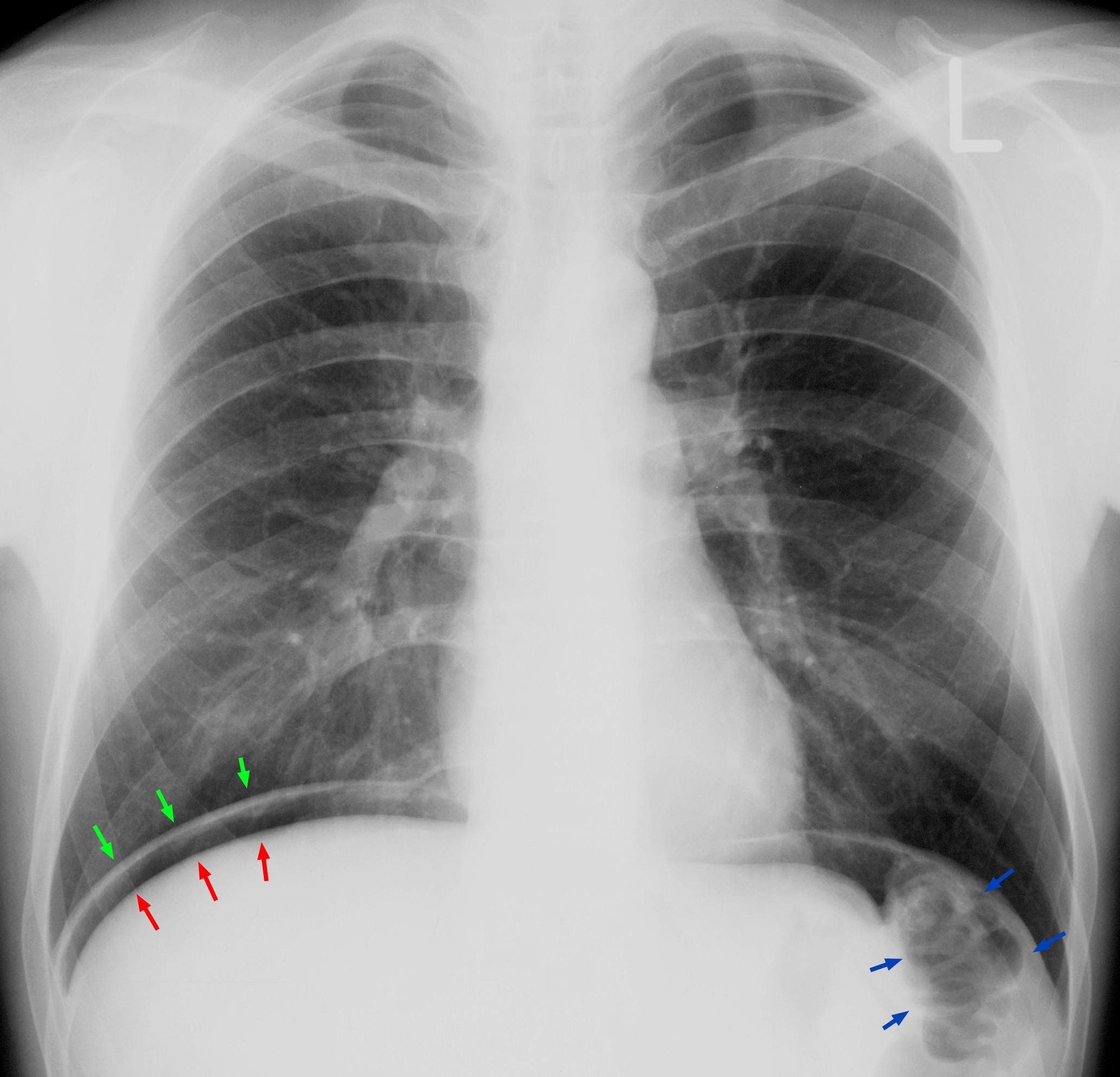
Diagnosing pneumoperitoneum is a piece of cake when you see the classic sign " Air under the diaphragm".
Air under diaphragm

But this sign is present in an erect x-ray. Suppose your patient is too sick to get an erect x-ray done ( might be incubated), and instead you get a supine film done, then these are the signs you should look out for.

Sunday, May 5, 2013

Mnemonic for today

LARP:
Relationship of the vagus nerves at the Gastroesophageal junction. 
Here the left vagus is anterior, and the right vagus is posterior.

Wednesday, September 9, 2009

AIIMS 2006(from AIPPG website)

LInk to original source-AIPPG.


Conjugated hyperbilirubinemia is seen in:

A. Gilbert’s syndrome

B. Griggler Najjar syndrome

C. Breast milk jaundice

D. Dubin Johnson syndrome

Ans. (D) Dubin Johnson syndrome

(Ref: Nelson’s Textbook of Pediatrics 17th Ed, Ch. 338, P-1321)

A 15-year-old female presented to the emergency department with history of recurrent epistaxis, hematuria and hematochezia. There was a history of profuse bleeding from the umbilicus stump at birth. Previous investigations revealed normal prothrombin time, activated partial thromboplastin time, thrombin time and fibrinogen levels. Her platelet counts as well as platelet function tests were normal but urea c1ot Jt. positive. Which one of the following clotting factor is most likely to be deficient?

A. Factor X

B. Factor XI

C. Factor XII

D. Factor XIII

Ans. (D) Factor XIII

(Ref: Nelson Pediatrics 17th Ed/P-1661)

Which one of the following is the characteristic feature of juvenile myoclonic epilepsy?

A. Myoclonic seizures frequently occur in morning

B. Complete remission is common

C. Response to anticonvulsants is poor

D. Associated absence seizures are present in majority of patients

Ans. (A) Myoclonic seizures frequently occur in the morning

(Ref: Harrison’s Principles of Internal Medicine 16th Ed. 2005—Part XV-Neurologic Disorders;Sec. 2-Diseases of the Central Nervous System;Ch. 348-Seizures and Epilepsy)

Plethoric lung fields are seen in all of the following conditions, except:

A. Atrial septal defect (ASD)

B. TAPVC (Total Anomalous Pulmonary venous connection)

C. Ebstein’s anomaly

D. Ventricular septal defect

Ans. (C) Ebstein’s anomaly

(Ref: Review of Radiology 3rd Ed/p-42-43)

Which of the following is an example of disorders of sex chromosomes?

A. Marfan’s syndrome

B. Testicular feminization syndrome

C. Klinefelter’ s syndrome

D. Down’s syndrome

Ans. (C) Klinefelter’ s syndrome

(Ref: Robbins and Cotrans’s Pathologic Basis of Disease 7th Ed/P-145)

Which of the following haemoglobin (Hb) estimation will be diagnostically helpful in a case of beta thalassemia trait?

A. Hb-F B. Hb1C

C. Hb-A2 D. Hb-H

Ans. (C) Hb-A2

(Ref: Nelsons Pediatrics 17th Ed/P-1633)

Which of the following circulating antibodies has the best sensitivity and specificity for the diagnosis of celiac disease?

A. Anti-endomysial antibody

B. Anti-tissue transglutaminase antibody

C. Anti-gliadin antibody

D. Anti-reticulin antibody

Ans. (A) Anti-endomysial antibody

(Ref: Nelsons Pediatrics 17th Ed/P-1265)

A couple has two children affected with tuberous sclerosis. On detailed clinical and laboratory evaluation (including molecular studies) both parents are normal. Which one of the following explains the two affected children in this family?

A. Non penetrance

B. Uniparental diasomy

C. Genomic imprinting

D. Germline mosaicism

Ans. (D) Germline Mosaicism

(Ref: Journal of Child Neurology/Vol. 19, No. 9, Sept. 2004)

Cardiomyopathy may be seen in all of the following except:

A. Duchenne muscular dystrophy

B. Friedreich’s ataxia

C. Type II glycogen storage disease

D. Alkaptonuria

Ans. (D) Alkaptonuria

Enzyme replacement therapy is available for which of the following disorders?

A. Gaucher disease

B. Niemann Pick disease

C. Mucolipidosis

D. Metachromatic leukodystrophy

Ans. (A) Gaucher’s disease (repeat)

In a child with acute liver failure, the most important prognostic factor for death is:

A. Increasing transaminases

B. Increasing bilirubin

C. Increasing prothrombin time

D. Gram negative sepsis

Ans. (C) Prothrombin time

(Ref: Diseases of the liver and the biliary system 11th Ed, Ch. 8-Acute Liver Failure, P-118)

Which of the following does not establish a diagnosis of congenital CMV infection in a neonate?

A. Urine culture of CMV

B. IgG CMV antibodies in blood

C. Intra-nuclear inclusion bodies in hepatocytes

D. CMV viral DNA in blood by polymerase chain reaction

Ans (B) IgG CMV antibodies in blood

(Ref: Cloherty’s Manual of Neonatal Care 5th Ed/P-257)

All of the following are true of β thalassemia major, except:

A. Splenomegaly

B. Target cells on peripheral smear

C. Microcytic hypochromic anemia

D. Increased osmotic fragility

Ans. (D) Increased osmotic fragility

(Ref: Manual of Pediatric Hematology and Oncology, 4th Ed/P-184)

Transient synovitis (toxic synovitis) of the hip is characterized by all of the following, except:

A. May follow upper respiratory infection

B. ESR and white blood cell counts are usually normal

C. Ultrasound of the joint reveals widening of the joint space

D. The hip is typically held in adduction and internal rotation

Ans. (D) The hip is typically held in adduction and internal rotation.

(Ref: Nelson’s Textbook of Pediatrics 17th Ed, Ch. 148, P-809)
A 3-year-old boy presents with fever, dysuria and gross hematuria. Physical examination shows a prominent suprapubic area which is dull to percussion. Urinalysis reveals red blood cells but no proteinuria. Which of the following is the most likely diagnosis?

A. Acute glomerulonephritis

B. Urinary tract infection

C. Posterior urethral valves

D. Teratoma

Ans. (B) Urinary tract infection

Which of the following statements is true of primary grade IV-V vesicoureteric reflux in young children?

A. Renal scarring usually begins in the midpolar regions

B. Postnatal scarring may occur even in the absence of urinary tract infections

C. Long-term outcome is comparable in patients treated with either antibiotic prophylaxis or surgery

D. Oral amoxicillin is the choice antibiotic for prophylaxis

Ans. (B) Postnatal scarring may occur even in the absence of urinary tract infections.

(Ref: Nelson’s Textbook of Pediatrics 17th Ed—Ch. 531-Vesicoureteric Reflux, P-1791-1793)

15-year-old boy presented with one day history of bleeding gums, subconjunctival bleed and purpuric rash. Investigations revealed the following results:

Hb-6.4 gm/dL; TLC-26,500/mm3 Platelet-35,000/mm3; prathrombin time–20 sec with a control of 13 sec; partial thromboplastin time-50 sec; and Fibrinogen 10 mg/dL. Peripheral smear was suggestive of acute myeloblastic leukernice. Which of the following is the most likely?

A. Myeloblastic leukemia without maturation

B. Myeloblastic leukemia with maturation

C. Promyelocytic leukemia

D. Myelomonscytic leukemia

Ans. (C) Promyelocytic leukemia

(Ref: Manual of Pediatric Hematology and Oncology, 4th Ed/P-306, 443)

The defective migration of neural crest cells results in:

A. Congenital megacolon

B. Albinism

C. Adrenogenital hypoplasia

D. Dentinogenesis imperfecta

Ans. (A) Congenital megacolon

(Ref: Schwartz’s Principles of Surgery 8th Ed. 2005— Part II- Specific Considerations; Ch. 38-Pediatric Surgery)

A premature infant is born with a patent ductus arteriosus. Its closure can be stimulated by administration of:

A. Prostaglandin analogue

B. Estrogen

C. Anti-estrogen compounds

D. Prostaglandin inhibitors

Ans. (D) Prostaglandin inhibitors

The loading dose of Aminophylline is:

A. 50-75 ug/kg

B. 0.5-1.0 mg/kg

C. 2.0-3.5 mg/kg

D. 5-6 mg/kg

Ans. (D) 5-6 mg/kg

Cushing’s Triad includes all except:

A. Hypertension

B. Bradycardia

C. Hypothermia

D. Irregular respiration

Ans. (C) Hypothermia

(Ref: Current Pediatric Diagnosis and Treatment 17th Ed. 2005—Ch. 11-Emergencies and Injuries)

All of the following drugs are used for managing status epilepticus except:

A. Phenytoin

B. Diazepam

C. Thiopentone sodium

D. Carbamazepine

Ans. (D) Carbamazepine

(Ref: Current Pediatric Diagnosis and Treatment 17th Ed. 2005—Ch. 23-Neurologic and Muscular Disorders; Table 23.9-Status epilepticus treatment)

Administration of glucose solution is prescribed for all of the following situations except:

A. Neonates

B. Child of a diabetic mother

C. History of unconsciousness

D. History of hypoglycemia

Ans. (C) History of unconsciousness

(Ref: Rudolph’s Pediatrics 21st Ed. 2003—24. The Endocrine System; 24.9-Hypoglycemia)

Which organ is the primary site of hematopoiesis in the fetus before midpregnancy?

A. Bone

B. Liver

C. Spleen

D. Lung

Ans. (B) Liver

(Ref: Nelson’s Textbook of Pediatrics 17th Ed/
P-1599)

All of the following are the complications in the new born of a diabetic mother except:

A. Hyper bilirubinemia

B. Hyperglycemia

C. Hypocalcemia

D. Hypomagnesemia

Ans. (B) Hyperglycemia

(Ref: Cloherty’s Manual of Neonatal Care 5th Ed./P-13-1
Sunday, August 16, 2009

Eponymous Signs in Splenic rupture

Ballance's  sign - Ballance's sign is dullness to percussion in the left flank LUQ and shifting dullness to percussion in the right flank seen with splenic rupture/hematoma. The dullness in the left flank is due to coagulated blood, the shifting dullness on the right due to fluid blood.

Kehr's  sign - Kehr's sign is the occurrence of acute pain in the tip of the shoulder due to the presence of blood or other irritants in the peritoneal cavity when a person is lying down and the legs are elevated. Kehr's sign in the left shoulder is considered a classical symptom of a ruptured spleen. Kehr's sign is a classical example of referred pain: irritation of the diaphragm is signalled by the phrenic nerve as pain in the area above the collarbone.

Saegasser's  sign  - Palpation of  left upper quadrant  inferior to ribs  elicits neck pain in the  patient. 

Friday, July 24, 2009

Different Eponymous types of Hernia

Amyand's hernia :The term Amyand’s hernia refers to the presence of the appendix within the hernial sac, and has been variously defined as the occurrence of either an inflammed or perforated appendix within an inguinal hernia, or simply, the presence of a non-inflammed appendix within an irreducible inguinal hernia.

The pathophysiology of Amyand’s hernia is unknown. Weber et al [4], proposed that appendix in
hernia becomes inflamed as a result of repeated trauma,leading to adhesions and bacterial overgrowth.

Barth's hernia :Hernia of the loops of intestine between the serosa of the abdominal wall and that of a persistent vitelline duct.

Beclard's hernia - femoral hernia through saphenous opening

Berger's hernia - hernia in Pouch of Douglas

Bochdalek hernia :(congenital posterolateral hernia of the diaphragm)A Bochdalek Hernia is one of two forms of a congenital diaphragmatic hernia, the other form being Morgagni's hernia.

The foramen of Bochdalek is a 2cm x 3cm opening in the posterior aspect of the diaphragm in the foetus, through which the pleuroperitoneal canal communicates between the pleural and peritoneal cavities. This canal normally closes by the 8thweek of gestation, failure or  incomplete fusion of the lateral (costal) with the posterior (crural) components of the diaphragm leads to the
development of Bochdalek hernia. Since the left canal closes later than the right, this type of hernia is found on the left side in 85% of cases

Cloquet's hernia :A femoral hernia perforating the aponeurosis of the pectineus and insinuating itself between this aponeurosis and the muscle, lying therefore behind the femoral vessels.


Cooper's hernia (bilocular femoral hernia ): A femoral hernia with two sacs, the first being in the femoral canal, and the second passing through a defect in the superficial fascia and appearing immediately beneath the skin.

De Garengeot's hernia : incarceration of the vermiform appendix within a femoral hernia.

Gibbon's hernia : hernia with hydrocoele

Gruber's hernia :Internal mesogastric hernia.

Hesselbach's hernia - hernia of a loop of intestine through the cribriform fascia presenting lateral to femoral artery

Hey's hernia :encysted hernia, scrotal or oblique inguinal hernia in which the bowel, enveloped in its own proper sac, passes into the tunica vaginalis in such a way that the bowel has three coverings of peritoneum

Holthouse hernia :an inguinal hernia that has turned outward into the groin.

Krönlein's hernia: An inguinoproperitoneal hernia; a hernia that is partially inguinal and partly properitoneal.

Larrey's hernia = (Morgagni's hernia)

Laugier’s femoral hernia- This is a type of femoral hernia through a gap in the lacunar ligament. It is more medial in position and nearly always strangulated.

Littre's hernia - hernia with Meckels's Diverticulum

lumbar hernia: hernia in the lumbar region (not to be confused with a lumbar disc hernia), contains following entities:

  • Petit's hernia - hernia through Petit's triangle (inferior lumbar triangle).
  • Grynfeltt's hernia - hernia through Grynfeltt-Lesshaft triangle (superior lumbar triangle).

Maydl's hernia -(hernia-in-W) The hernia contains two loops of bowel arranged like a 'W'. The central loop of the 'W' lies free in the abdomen and is strangulated where as the two loops present in the sac are not.

Mesocolic / transmesenteric hernias:  occur through iatrogenically created defects in the mesentery. These defects include herniation of an abdominal viscus, usually through the small bowel mesentery or transverse mesocolon. These hernias are common following abdominal surgery, especially Roux-en-Y loop reconstruction,  which creates a defect in the mesentery.

Morgagni hernia (also known as retrosternal or parasternal diaphragmatic hernia) occurs due to the defective fusion of the septal transverses of the diaphragm and the costal arches. This anatomic defect lies posterolateral to the sternum and is called Larrey’s space . The exact aetiology of this hernia is unknown but it is postulated that it begins as a weakness in the diaphragm which is later stretched due to intraperitoneal pressure.

Narath’s femoral hernia - The hernia lies hidden behind the femoral vessels. It occurs only in patients with congenital hip dislocation due to lateral displacement of the psoas muscle.

Pantaloon hernia: a combined direct and indirect hernia, when the hernial sac protrudes on either side of the inferior epigastric vessels

Perineal hernia(Mery’s hernia): A perineal hernia protrudes through the muscles and fascia of the perineal floor. It may be primary but usually, is acquired following perineal prostatectomy, abdominoperineal resection of the rectum, or pelvic exenteration.

Phantom hernia - Localised muscle buldge following muscular paralysis

Richter's hernia: strangulated hernia involving only one sidewall of the bowel, which can result in bowel perforation through ischaemia without causing bowel obstruction or any of its warning signs.

Rieux's hernia :retrocecal hernia, protrusion of the intestine into a pouch behind the cecum.

Rokitansky's hernia :A separation of the muscular fibres of the bowel allowing protrusion of a sac of the mucous membrane.

Serofini's hernia - behind femoral vessels

Spigelian hernia - Spigelian hernia occurs through congenital or acquired defects in the spigelian fascia. This is the area of the transversus abdominis aponeurosis, lateral to the edge of the rectus muscle but medial to the spigelian line, which is the point of transition of the transversus abdominis muscle to its aponeurotic tendon.

Treitz's hernia is the eponymous name for a paraduodenal hernia. These are rare hernias that arise in the potential spaces and folds of the posterior parietal peritoneum adjacent to the ligament of Treitz.(duodenojejunal hernia)

Velpeau hernia: A velpeau hernia is a femoral hernia in front of the femoral blood vessels in the groin.


image

Thursday, July 9, 2009

Eponyms for Percivall Pott


Eponym Description
Pott's aneurysm Arteriovenous aneurysm in which blood flows from an artery directly into a vein without going through a connecting sac
Pott's cancer Scrotal cancer originally described in chimney sweeps exposed to coal dust
Pott's disease Caries or osteitis of the vertebrae, usually of tuberculous origin (mycobacterium tuberculosis), characterized by softening and collapse of the vertebrae, often resulting in kyphosis, a hunchback deformity (Pott's curvature).
Pott's fracture Supramalleolar fracture of 1 or both ankle bones
Pott's gangrene Gangrene of foot from arterial insufficiency
Pott's paraplegia Paraplegia caused by spinal cord compression and abscesses in tuberculous spondylitis (Pott disease)
Pott's puffy tumor Pott puffy tumor is a subperiosteal abscess of the frontal bone that appears as a localized swelling of the overlying region of the forehead. It is the result of frontal sinusitis and osteomyelitis that erode through the anterior mantle of the frontal bone.

Wednesday, July 8, 2009

Splitting of Second heart sound

To understand Splitting of S2 better, we should first understand its normal physiology.

Second heart sound:

It has two audible components, the aortic valve closure sound (A2) and the pulmonic valve closure sound (P2), which are normally split on inspiration and virtually single on expiration.

Technique:

It has two audible components, the aortic closure sound (A2) and the pulmonic closure sound (P2), which must be separated by more than 20 msec (0.20 sec) in order to be differentiated and heard as two distinct sounds. It is clinically very important to determine the presence and degree of respiratory splitting and the relative intensities of A2 and P2.

Splitting is best identified in the second or third left ICS, since the softer P2 normally is confined to that area, whereas the louder A2 is heard over the entire precordium, including the apex.

Physiology:

Various research findings suggests that closure of the aortic and pulmonic valves initiates the series of events that produces the second heart sound. The main audible components, however, result from vibrations of the cardiac structures after valve closure. Using high-fidelity, catheter-tipped micro-manometers and echophonocardiography, it has been shown that the aortic and pulmonic valves close silently and that co-aptation of the aortic valve cusps precedes the onset of the second sound by a few milliseconds. The second sound therefore originates from after-vibrations in the cusps and in the walls and blood columns of the great vessels and their respective ventricles. The energy from these oscillations comes from sudden deceleration of retrograde flow of the column of blood in the aorta and pulmonary artery when the elastic limits of the tensed valve leaflets are met. This abrupt deceleration sets the whole cardiohemic system into vibration.

In order to understand splitting of the second heart sound, knowledge of its relationship to the cardiac cycle is essential.

First lets understand why is A2 before P2 ?

1.Both right & left ventricular systole ends at the same time .(ie, systolic muscle contraction of both ventricles ends at same time)

2.pulmonary arterial pressure is less than aortic pressure (i.e, pulmonary resistance to forward flow from ventricles is less than aortic resistance => therefore we can say that pulmonary impedance is less than aortic impedance.){impedance is nothing but resistance}

3.Therefore as pulmonary impedance is less, even after right ventricular systolic contraction blood continues to flow through valve until pulmonary arterial pressure increases more than right ventricle). But as aortic impedance is more ,it stops blood flow through the aortic valve before itself.

4.Due to the above reasons ,Right ventricular ejection begins prior to left ventricular ejection, has a slightly longer duration, and terminates after left ventricular ejection, resulting in P2 normally occurring after A2.

A2 and P2 are coincident with the incisura of the aorta and pulmonary artery pressure curves, respectively, and terminate left and right ventricular ejection periods.(incisura reflects closure of valves)

The differences between the aortic and pulmonary artery vascular impedance characteristics are also essential to understanding the effects of respiration on splitting of S2. When the pressure curves of the pulmonary artery and right ventricle are recorded simultaneously, the pulmonary artery curve at the level of the incisura (dicrotic notch) lags behind the right ventricular curve, or "hangs out" after it.

hangout interval

The duration of the "hangout interval" is a measure of impedance in the pulmonary artery system. In the highly compliant (low-resistance, high-capacitance) pulmonary vascular bed, the hangout interval may vary from 30 to 120 msec, contributing significantly to the duration of right ventricular ejection.

In the left side of the heart, because impedance is much greater, the hangout interval between the aorta and left ventricular pressure curves is negligible (less than or equal to 5 msec).

The hangout interval therefore correlates closely with impedance of the vascular bed into which blood is being injected. Its duration appears to be inversely related to vascular impedance.

Normal physiological splitting during respiration:

Alterations in the impedance characteristics of the pulmonary vascular bed and the right-sided hangout interval are responsible for many of the observed changes in splitting of S2.

In a normal physiologic setting, inspiration lowers impedance in the pulmonary circuit, prolongs the hangout interval and delays pulmonic valve closure, resulting in audible splitting of A2 and P2.

On expiration, the reverse occurs: pulmonic valve closure is earlier, and the A2–P2 interval is separated by less than 30 msec and may sound single to the ear. Since the pulmonary circulation has a much lower impedance than the systemic circulation, flow through the pulmonic valve takes longer than flow through the aortic valve. The inspiratory split widens mainly because of delay in the pulmonic component.

Traditionally it was believed that an inspiratory drop in intrathoracic pressure favored greater venous return to the right ventricle, pooling of blood in the lungs, and decreased return to the left ventricle. The increase in right ventricular volume prolonged right-sided ejection time and delayed P2; the decrease in left ventricular volume reduced left-sided ejection time and caused A2 to occur earlier. The delayed P2 and early A2 associated with inspiration, however, are best understood as an interplay between changes in the pulmonary vascular impedance and changes in systemic and pulmonary venous return. The net effect is that right ventricular ejection is prolonged, left ventricular ejection is shortened, and the A2–P2 interval widens during inspiration.

Clinical Significance

Normally the aortic closure sound (A2) occurs prior to the pulmonic closure sound (P2), and the interval between the two (splitting) widens on inspiration and narrows on expiration. With quiet respiration, A2 will normally precede P2 by 0.02 to 0.08 second (mean, 0.03 to 0.04 sec) with inspiration. In younger subjects inspiratory splitting averages 0.04 to 0.05 second during quiet respiration. With expiration, A2 and P2 may be superimposed and are rarely split as much as 0.04 second. If the second sound is split by greater than 0.04 second on expiration, it is usually abnormal.

Therefore, the presence of audible splitting during expiration (i.e., the ability to hear two distinct sounds during expiration) is of greater significance at the bedside in identifying underlying cardiac pathology than is the absolute inspiratory increase in the A2–P2 interval.

ABNORMAL SPLITTING OF THE SECOND HEART:

(1) persistently single;

(2) persistent (audible expiratory) splitting, with normal respiratory variation;

(3) persistent splitting without respiratory variation (fixed splitting); and

(4) reversed (paradoxical) splitting.

splitting s2

(1) persistently single;

  • When S2 remains single throughout the respiratory cycle, one component is absent or the two components are persistently synchronous.
  • The most common cause of a single S2 is inaudibility of the P2 in older adults with increased anteroposterior chest dimensions.
  • In the setting of
    congenital heart disease, a single S2 due to absence of the pulmonary component is a feature of pulmonary atresia, severe pulmonary valve stenosis, dysplastic pulmonary valve, or complete transposition of the great arteries.
  • Conversely, a single S2 due to inaudibility of the A2 occurs when the aortic valve is immobile (severe calcific aortic stenosis) or atretic (aortic atresia).

(2) persistent (audible expiratory) splitting, with normal respiratory variation;

  • Persistent splitting may be due to a delay in P2, as in cases of simple complete right bundle branch block,or to early timing of the A2, as occasionally occurs in cases of mitral regurgitation(since early emptying of left ventricle –> early closure of Aortic valve).
  • Normal directional changes in the interval of the split (greater with inspiration, lesser with exhalation) in the presence of persistent audibility of both components defines the split as persistent but not fixed.
  • these conditions have wide inspiratory split

(3) fixed splitting;

  • This term applies when the interval between the A2 and P2 is not only wide and persistent but also remains unchanged during the respiratory cycle.
    Fixed splitting is an auscultatory hallmark of  atrial septal defect(ASD). 
  • A2 and P2 are widely separated during exhalation and exhibit little or no change in the degree of splitting during inspiration.

Reason behind wide splitting:

The wide splitting is caused by a delay in the P2 because a marked decrease in pulmonary vascular impedance prolongs the interval between the descending limbs of the pulmonary arterial and right ventricular pressure pulses (“hangout”), and therefore delays the pulmonary incisura and the P2.

Reason behind fixed splitting:

  • We know that in normal individuals , the amount of pulmonary ejection is more during inspiration compared to that in expiration (since pulmoary impedance decreases during inspiration)
  • In ASD .the amount of blood which passes throught he pulmonary valve ramains the samee in both inspiration& expiration because of Phasic changes in systemic venous return during respiration in patients with atrial septal defect are associated with reciprocal changes in the volume of the left-to-right shunt, minimizing respiratory variations in right ventricular filling. (ie, whenever venous return increases in inspiration, it causes a reciprocal decrease in left to right shunting in atria & whenever venous return decreased as in expiration ,shunting increases =>this maintains equal amount of blood in right ventricle irrespective of inspiration or exoiration)
  • The net effect is the characteristic wide, fixed splitting of the two components of the S2.

(4) reversed (paradoxical) splitting. 

  • This term refers to a reversed sequence of semilunar valve closure, the P2 preceding the A2.
  • Common causes of paradoxical splitting are complete left bundle branch block or a right ventricular pacemaker, both of which are associated with initial activation of the right side of the ventricular septum, and delayed activation of the left ventricle owing to transseptal (right-to-left) depolarization.
  • When the S2 splits paradoxically, its two components separate during exhalation and become
    single (synchronous) during inspiration .
  • Inspiratory synchrony is achieved as the two components fuse because of a delay in the P2, less to earlier timing of the aortic
    component.

  splitting s2


Please review this article & if you have any doubts do tell me.

Thursday, July 2, 2009

Brainstem Rules of 4


In 2005, Peter Gates published a superb paper titled:

‘The rule of 4 of the brainstem: a simplified method for understanding brainstem anatomy and brainstem vascular syndromes for the non-neurologist’.

Gates described a simplified method for answering the question ‘Where is the lesion?’ using only the parts of the brainstem that we actually examine during a clinical examination to understand brainstem vascular syndromes.

Firstly, a quick review of the blood supply of the brainstem. Simply put the blood supply comes from:

  1. paramedian branches
  2. long circumferential branches (SAP)
    • superior cerebellar artery (SCA)
    • anterior inferior cerebellar artery (AICA)
    • posterior inferior cerebellar artery (PICA)

And occlusion of these two groups of vessels results in two distinct types of brainstem syndrome:

  1. medial (or paramedian) brainstem syndromes ( due to para-median branch occlusion)
  2. lateral brainstem syndromes ( due to occlusion of the circumferential branches, also occasionally seen in unilateral vertebral occlusion) blood2

And now the rules. If you can remember these rules the  diagnosis of brainstem vascular syndromes becomes a pitifully simple exercise (?!) - here’s how it works:

In the rule of 4 there are 4 rules

  1. There are 4 structures in the ‘midline‘ beginning with M
  2. There are 4 structures to the ‘side‘ (lateral) beginning with S
  3. There are 4 cranial nerves in the medulla, 4 in the pons and 4 above the pons (2 in the midbrain)
  4. The 4 motor nuclei that are in the midline are those that divide equally into 12 except for 1 and 2, that is 3, 4, 6 and 12
    (5, 7, 9 and 11 are in the lateral brainstem)

The 4 medial structures and the associated deficits are:

  1. Motor pathway (or corticospinal tract):
    contralateral weakness of the arm and leg
  2. Medial Lemniscus:
    contralateral loss of vibration and proprioception in the arm and leg
  3. Medial longitudinal fasciculus:
    ipsilateral inter-nuclear ophthalmoplegia
    (failure of adduction of the ipsilateral eye towards the nose and nystagmus in the opposite eye as it looks laterally)
  4. Motor nucleus and nerve:
    ipsilateral loss of the cranial nerve that is affected (3, 4, 6 or 12)

The 4 ’side’ (lateral) structures and the associated deficits are:

  1. Spinocerebellar pathway:
    ipsilateral ataxia of the arm and leg
  2. Spinothalamic pathway:
    contralateral alteration of pain and temperature affecting the arm, leg and rarely the trunk
  3. Sensory nucleus of the 5th cranial nerve:
    ipsilateral alteration of pain and temperature on the face in the distribution of the 5th cranial nerve
    (this nucleus is a long vertical structure that extends in the lateral aspect of the pons down into the medulla)
  4. Sympathetic pathway:
    ipsilateral Homer’s syndrome, that is partial ptosis and a small pupil (miosis)

According to Gates:

These pathways pass through the entire length of the brainstem and can be likened to ‘meridians of longitude‘ whereas the various cranial nerves can be regarded as ‘parallels of latitude‘. If you establish where the meridians of longitude and parallels of latitude intersect then you have established the site of the lesion.

The 4 cranial nerves in the medulla are CN9-12:

  1. Glossopharyngeal (CN9):
    ipsilateral loss of pharyngeal sensation
  2. Vagus (CN10):
    ipsilateral palatal weakness
  3. Spinal accessory (CN11):
    ipsilateral weakness of the trapezius and stemocleidomastoid muscles
  4. Hypoglossal (CN12):
    ipsilateral weakness of the tongue

The 12th cranial nerve is the motor nerve in the midline of the medulla. Although the 9th, 10th and 11th cranial nerves have motor components, they do not divide evenly into 12 (using our rule) and are thus not the medial motor nerves.

The 4 cranial nerves in the pons are CN5-8:

  1. Trigeminal (CN5):
    ipsilateral alteration of pain, temperature and light touch on the face back as far as the anterior two-thirds of the scalp and sparing the angle of the jaw.
  2. Abducent (CN6):
    ipsilateral weakness of abduction (lateral movement) of the eye (lateral rectus).
  3. Facial (CN7):
    ipsilateral facial weakness.
  4. Auditory (CN8):
    ipsilateral deafness.

The 6th cranial nerve is the motor nerve in the medial pons.
The 7th is a motor nerve but it also carries pathways of taste, and using the rule of 4 it does not divide equally in to 12 and thus it is not a motor nerve that is in the midline.
The vestibular portion of the 8th nerve is not included in order to keep the concept simple and to avoid confusion. Nausea and vomiting and vertigo are often more common with involvement of the vestibular connections in the lateral medulla.

The 4 cranial nerves above the pons are CN1-4:

  1. Olfactory (CN1):
    not in midbrain.
  2. Optic (CN2):
    not in midbrain.
  3. Oculomotor (CN3):
    impaired adduction, supradduction and infradduction of the ipsilateral eye with or without a dilated pupil.
    The eye is turned out and slightly down.
  4. Trochlear (CN4):
    eye unable to look down when the eye is looking in towards the nose (superior oblique).

The 3rd and 4th cranial nerves are the motor nerves in the midbrain.

Thus a medial brainstem syndrome will consist of the 4 M’s and the relevant motor cranial nerves, and a lateral brainstem syndrome will consist of the 4 S’s and either
the 9-11th cranial nerve if the lesion is in the medulla, or the 5th, 7th and 8th cranial nerve if the lesion is in the pons.

Handy tip:
If there are signs of both a lateral and a medial (paramedian) brainstem syndrome, then one needs to consider a basilar artery problem, possibly an occlusion.

I’ll let you mull over these rules until the next ‘brainstem’ post, where you’ll be able to test drive ‘Gates’ Brainstem Rules of 4′ on some clinical scenarios.

Scenario 1

  • You are examining a patient with sudden onset left-sided weakness. These are your clinical examination findings:
    weakness of the left upper and lower limbs, with sparing of the face.
    tongue deviation to the right, with no ophthalmoplegia.
    loss of vibration and proprioception in the left upper and lower limbs.
  • medial_brainstem_synd
  • Where is the lesion?
    weakness of the left upper and lower limbs, with sparing of the face:
    motor (corticospinal pathway) localises the lesion to the contralateral medial brainstem
    (sparing of the face (CN7) means the lesion must be below the upper pons)
    tongue deviation to the right, with no ophthalmoplegia:
    tongue deviation indicates CN12 involvement, localising the lesion to the ipsilateral medulla
    (sparing of CN3 and CN6 means the midbrain and pons are not involved)
    loss of vibration and proprioception in the left upper and lower limbs:
    confirms localisation of the lesion to the contralateral medial brainstem
  • Site of the lesion: right medial medulla. Sometimes, due to the peculiar pattern of blood supply to the medulla, bilateral infarction may occur.

Scenario 2

  • You are examining a patient with sudden onset right-sided weakness. These are your clinical examination findings:
    weakness of the right face, upper and lower limbs.
    the left eye is turned “down and out” and the pupil is dilated.
  • Where is the lesion?
    weakness of the right face, upper and lower limbs:
    motor (corticospinal pathway) localises the lesion to the contralateral medial brainstem
    (involvement of the face means the lesion must be at or above the upper pons)
    the left eye is turned “down and out” and the pupil is dilated:
    CN3 involvement, localising the lesion to the ipsilateral midbrain
    (sparing of CN6 and CN12 means the pons and medulla are not involved)
  • Site of the lesion: left medial midbrain. A CN3 palsy (from damage to the CN3 nerve fascicle) and contralateral hemiplegia is known as Weber’s syndrome (”basal” infarction)  - which can be difficult to distinguish from ‘coning’ if you don’t have a CT scanner available.

Scenario 3

  • You are examining a patient with vertigo, vomiting, and nystagmus. These are your clinical examination findings:
    left-sided limb ataxia.
    left-sided alteration of pain and temperature on the face.
    left-sided ipsilateral Homer’s syndrome.
    right-sided alteration of pain and temperature affecting the arm and leg.
    dysarthria and decreased gag reflex on the left, with the palate pulling up on the right-side.
  • wallenberg syndrome
  • Where is the lesion?
    left-sided limb ataxia:
    spinocerebellar pathway localises the lesion to the ipsilateral lateral brainstem.
    left-sided alteration of pain and temperature on the face:
    Sensory nucleus of the 5th cranial nerve localises the lesion to the ipsilateral lateral brainstem.
    left-sided ipsilateral Homer’s syndrome:
    Sympathetic pathway localises the lesion to the ipsilateral lateral brainstem.
    right-sided alteration of pain and temperature affecting the arm and leg:
    Spinothalamic pathway localises the lesion to the contralateral lateral brainstem.
    dysarthria and decreased gag reflex on the left, with the palate pulling up on the right-side:
    localises the lesion to the medulla affecting the ipsilateral  CN9 and 10.
  • Site of the lesion: left lateral medulla. Also known as Wallenberg’s syndrome, caused by a left vertebral or left posterior inferior cerebellar artery occlusion (blood supply is variable to this region).

Scenario 4

  • You are examining a patient with right-sided deafness, that was preceded by tinnitus. These are your clinical examination findings:
    right-sided limb ataxia (predominantly affecting the right upper limb).
    right-sided facial numbness with loss of the corneal reflex.
    right-sided hemi-facial spasms.
  • Where is the lesion?
    right-sided limb ataxia (predominantly affecting the right upper limb):
    spinocerebellar pathway localises the lesion to the ipsilateral lateral brainstem.
    right-sided facial numbness with loss of the corneal reflex:
    Sensory nucleus of the 5th cranial nerve localises the lesion to the ipsilateral lateral brainstem.
    right-sided hemi-facial spasms:
    the lesion involves the pons affecting the ipsilateral CN7.
  • Site of the lesion: The findings indicate a lesion affecting the right lateral pons with evidence of spinocerebellar involvement. In this case the lesion was not vascular in origin but in fact an example of a cerebropontine angle lesion - an acoustic neuroma (or schwannoma). This demonstrates the the broader utility of Gates’ Brainstem Rules of 4.

Scenario 5

  • You are examining a patient with sudden onset right-sided weakness. These are your clinical examination findings:
    weakness of the right face, upper and lower limbs.
    failure of abduction of the left eye.
    loss of vibration and proprioception in the right upper and lower limbs.
  • Where is the lesion?
    weakness of the right face, upper and lower limbs:
    motor (corticospinal pathway) localises the lesion to the contralateral medial brainstem
    (involvement of the face means the lesion must be at or above the upper pons)
    failure of abduction of the left eye:
    indicates CN6 involvement, localising the lesion to the ipsilateral pons.
    (sparing of CN3 and CN12 means the midbrain and medulla are not involved)
    loss of vibration and proprioception in the right upper and lower limbs:
    confirms localisation of the lesion to the contralateral medial brainstem
  • Site of the lesion: left medial pons. Interestingly, the facial nerve runs a strange course - it loops around medial to the CN6 nucleus from its own laterally situated CN7 nucleus. Thus a CN7 palsy tends to coexist with a CN6 lesion despite the CN7 nucleus being in the lateral pons.

Scenario 6

  • You are examining a patient with sudden onset intermittent double vision (diplopia). These are your clinical examination findings:
    failure of adduction past the midline (movement towards the nose) of the left eye and leading eye (right) nystagmus on looking laterally to the right. Normal eye movements on looking to the left.
    The patient is hypertensive. There is no hemiparesis and further examination is unremarkable.
  • Where is the lesion?
    This finding  suggests a unilateral left-sided internuclear ophthalmoplegia, which localises the lesion to the ipsilateral medial longitudinal fasciculus (MLF). The MLF connects CN3 in the midbrain and the contralateral CN6 in the pons.
    The MLF is not usually affected when there is hemiparesis as it lies further back in the brainstem relative to the motor (corticospinal) pathway.
    Unilateral internuclear ophthalmoplegia can result from a lacunar infarct.
    (Always remember the possibility of multiple sclerosis)
  • Site of the lesion: left medial longitudinal fasciculus (connects CN3 in the midbrain and contralateral CN6 in the pons).

Scenario 7

  • You are examining a patient with a right-sided Horner’s syndrome. These are your clinical examination findings:
    right-sided Horner’s syndrome.
    right-sided limb ataxia.
    left-sided total loss of sensation.
  • Where is the lesion?
    right-sided Horner’s syndrome:
    sympathetic pathway localises the lesion to the ipsilateral lateral brainstem.
    right-sided limb ataxia:
    spinocerebellar pathway localises the lesion to the ipsilateral lateral brainstem.
    left-sided total loss of sensation:
    Spinothalamic pathway localises the lesion to the contralateral lateral brainstem (in the midbrain the medial lemniscal pathway is actually situated more laterally, ventral to the spinothalamic pathway - ie. the two pathways come together… an exception to the Rule of 4!).
  • Site of the lesion: Right dorsolateral midbrain. An extensive lesion that also involves CN3 is known by the delightful name of Nothnagel’s syndrome.

Scenario 8

  • You are examining a patient with a ‘down and out’ right eye with pupillary dilatation. These are your clinical examination findings:
    right-sided impaired adduction, supradduction and infradduction of the ipsilateral eye with a dilated pupil.
    left-sided limb ataxia.
  • Where is the lesion?
    right-sided impaired adduction, supradduction and infradduction of the ipsilateral eye with a dilated pupil:
    CN3 lesion localises the lesion to the ipsilateral medial midbrain.
    left-sided limb ataxia:
    usually this indicates ipsilateral spinocerebellar pathway involvment (a lateral structure). However, in this case we know that the midbrain is affected (CN3 palsy) and the red nucleus lies in the medial midbrain just lateral to the CN3 nerve fascicle. Damage to the red nucleus interrupts the ‘dentatorubrothalamic tract’ from the opposite cerebellar hemisphere causing cerebellar signs in the limbs opposite to the CN3 lesion.
  • Site of the lesion: Right medial midbrain. The clinical manifestations of this lesion (affecting the CN3 nucleus or its fascicle as well as the red nucleus) is known as Benedikt’s syndrome.


Taken from: Life in the Fast Lane

Wednesday, July 1, 2009

Clinical Image – What are they?

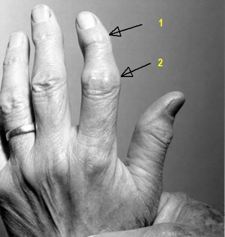
 heberden &bouchard nodes

1.Heberden’s nodes :

  • Heberden’s nodes are a clinical sign typically associated with osteoarthritis
  • Heberden’s nodes are often described as enlargements of the terminal (or distal) interphalangeal joints of the fingers
  • The histologic nature of the nodes is unknown.
  • They normally do not arise during the acute stage of osteoarthritic development, but rather during the chronic stage.In the acute stage, joints become erythematous, warm, and very tender to the touch. At this point, the joints are usually slightly swollen and may exhibit effusion. In addition, during the acute stage, pain in the area of the joints is often severe, occurring in spasms and with the sensation of burning and tingling in the skin overlying the joints. After several months, once the individual is in the chronic stage, the signs of inflammation disappear and bony outgrowths (or Heberden’s nodes) begin to arise over the terminal interphalangeal joints of the fingers. At this time, the joints are generally painless, and the characteristic deformation associated with Heberden’s nodes becomes obvious.

2.Bouchard's nodes :

  • Bouchard's nodes are comparable in presentation to Heberden's nodes, but are significantly less common.
  • They are hard, bony outgrowths on the proximal interphalangeal joints (the middle joints of fingers or toes.)
  • They are also a sign of osteoarthritis, and are caused by formation of bony outgrowths .

Sunday, June 28, 2009

Eponymous Signs of Aortic Regurgitation

Austin Flint Murmur

Description

The murmur typically begins in mid-diastole, often has a presystolic accentuation, and terminates at the end of diastole. It is low-pitched, with a rough and rumbling quality, and best heard at the apex. An Austin Flint murmur can be deemed present only in the setting of aortic regurgitation without coexisting mitral stenosis, since the latter can generate a similar murmur.

Pathophysiology

Austin Flint postulated that regurgitant blood flow in severe aortic regurgitation impinges on the leaflets of the mitral valve, leading to a functional stenosis. Diastolic inflow across this narrowed mitral valve orifice generates turbulence that is clinically appreciable as a mid- to late diastolic murmur . Later investigations have advanced a variety of other theories as causes for the murmur, including overlap of aortic regurgitation and mitral inflow jets , fluttering of mitral valve leaflets (, and left ventricular endocardial vibrations due to the aortic regurgitation jet. However, a universally accepted explanation remains elusive.

Elicitation

The murmur is best heard on auscultation at the apex by using the bell of the stethoscope, with the patient in the left lateral position.


Corrigan Pulse:

Description

Large volume collapsing pulse of the carotid artery

Pathophysiology

A recent investigation found that patients with aortic regurgitation had increased amplitude of the pulse, lower mean arterial pressure, and narrower pulse pressure than normal patients . The investigators concluded that these characteristics reflected an increase in the compliance of the arterial wall in patients with aortic regurgitation.

Elicitation

The examiner palpates the patient's radial artery while elevating the wrist. If the pulse clearly increases in amplitude, then the sign is present.


Duroziez Sign

Description

The sign denotes an intermittent to-and-fro femoral artery murmur (occurring in systole and diastole, respectively) generated by femoral artery compression.

Pathophysiology

Duroziez believed the systolic portion of the murmur was caused by forward flow into the lower extremity and that the diastolic segment was caused by aortic regurgitation toward the heart.

Elicitation

Duroziez auscultated the femoral artery while applying digital compression proximal and distal to the stethoscope .Blumgart and Ernstene  ) replaced digital compression with cephalad and caudad tilting of the stethoscope.


Hill Sign:This sign is also known as the popliteal–brachial gradient.

Description

A 20 mmHg difference in the popliteal and brachial systolic cuff pressures

Pathophysiology

The Hill sign, therefore, remains an unexplained artifact of indirect blood pressure measurement that is consistently more common and pronounced in patients with aortic regurgitation than in those without.

Elicitation

The blood pressure is manually obtained over the brachial and femoral arteries by using appropriately sized cuffs, with the patient in the recumbent position. The difference in systolic pressures denotes the gradient.


Watson’s water hammer pulse: also known as collapsing pulse, cannonball pulse or pulsus celer.

Description

Large-volume, ‘collapsing’ bounding peripheral pulses.

Pathogenesis        

The abrupt jerky, forceful upstroke of the whp implies a rapid filling of the radial artery in systole due to an extra large amount of blood pushed by the distended left ventricle into relatively empty arterial vessels. The collapsing or sudden down stroke may be partly due to a sudden fall in the diastolic pressure in the aorta due to regurgitation of blood into the left ventricle through a leaky valve and partly due to the rapid emptying of the arterial system due to the marked increase in the velocity of the bloodstream. Apart from the pathogenetic factors which cause the widened pulse pressure, lifting the patient's arm vertically upwards when eliciting the whp, helps the blood to empty quickly into the heart during diastole due to the gravity and also brings the radial artery more in line with the outflow stream of the aorta, thus accentuating the sign.


Traube’s sign/pistol shot sounds :

Booming systolic and diastolic sounds heard over the femoral artery when it is compressed distally

De Musset’s sign: Head nodding with each heart beat

Quincke’s sign :Pulsations in the nail capillary bed seen when light is transmitted through the fingertips or exerting gentle pressure on the tip of a fingernail .

Lighthouse sign:Blanching and flushing of forehead

Landolfi’s sign :Alternating constriction and dilatation of pupil

Becker’s sign :Visible pulsations of the retinal arterioles

Mueller’s sign :Visible pulsations of uvula

Mayen’s sign :Diastolic drop of BP >15 mmHg with arm raised

Rosenbach’s sign :Pulsatile liver

Lincoln sign :Pulsatile popliteal artery

Gerhardt’s sign : Pulsatile spleen

Sherman sign: Prominently located and palpated dorsalis pedis pulse

Thursday, June 25, 2009

Cardiovascular Examination – From Braunwauld’s Textbook

Braunwald 7 - Chapter 08 - Physical Examination

MEDMASTER FREE DOWNLOADS!

Download the below titles for free legally from MEDMASTER publications itself. Check this MEDMASTER Link

ATLAS OF MICROBIOLOGY

by Stephen Goldberg, M.D. A visual atlas of microorganisms, including viruses, bacteria, fungi, protozoans, helminths, prions, and the diseases they cause. An interactive computer program based on Internet searches for images and literature. A perfect complement to Gladwin: Clinical Microbiology Made Ridiculously Simple.
MEDSEARCHER

by Stephen Goldberg, M.D. A rapid way of searching through any one of the many key Internet medical search engines for instant medical information. Simply type in a topic and click "Search." Information appears within seconds.
ATLAS OF HUMAN DISEASES

by Stephen Goldberg, M.D. Explore the vast range of human diseases in this atlas of the many thousands of diseases that affect people, including photos and literature
ATLAS OF DERMATOLOGY

by Stephen Goldberg, M.D. and Barry Galitzer, M.D. A visual atlas of common and not-so-common dermatologic diseases. Accesses thousands of pictures via the Internet, along with literature related to diagnosis and treatment.   
USMLE STEP 2 QUESTION BANK

by Andreas Carl. A uniquely organized rapid question review for USMLE Step 2. Supplement to USMLE Step 2 Made Ridiculously Simple, by Andreas Carl. 1000+ questions.

ATLAS OF ORTHOPEDICS

by Patrice Tétreault, Hugue Ouellette, and Stephen Goldberg. An Internet-based atlas of clinical Orthopedics, with photos and movies corresponding to the text of Orthopedics Made Ridiculously Simple.     

Monday, June 22, 2009

Important Murmurs

Austin Flint murmur : Austin Flint murmur is a mid-diastolic rumbling audible in subjects with severe aortic regurgitation that is best heard at the apex with little radiation. Several theories have been suggested for the origin of the murmur: (1) vibration of the anterior mitral valve leaflet due to the regurgitant jet, (2) collision of the jet with mitral inflow, (3) increased mitral inflow velocity due to narrowing of the valve orifice by the jet, and (4) vibration from the jet impinging on the myocardial wall. It differs from mitral stenosis murmur in that ,it occurs in the presence of a murmur of aortic valve insufficiency and in the absence of the rheumatic, mitral opening snap.

Carey Coombs murmur : Mitral valvulitis associated with acute rheumatic fever may cause a low-pitched mid-diastolic rumble. It can be differentiated from the diastolic murmur of Mitral stenosis by the absence of (1)an opening snap, (2)presystolic accentuation & (3)loud first heart sound.

Cruveilhier-Baumgarten murmur : Venous hum heard in epigastric region (on examination by stethoscope) due to collateral connections between portal system and the remnant of the umbilical vein in portal hypertension.

Duroziez's murmur : Its a to & fro murmur heard over the femoral artery during both systole& diastole. It is elicited by applying gradual arterial compression with the diaphragm of the steth. This compression not only produces systolic murmur(which is the normal result of arterial compression) but also a diastolic murmur(which is pathologic & suggestive of aortic regurgitation).Sensitivity of 58-100%.False positives occur in high output states. In high output states the double murmur is due to forward flow. In Ar one murmur is due to forward flow& the other due to reverse flow.The two can be differnetiated by applying pressure first on the more cephalad edge of the diaphragm & then on its more caudal edge. The murmur of forward flow is enhanced by compressing the cephalad edge. Conversely the reverese flow murmur is enhanced by compressing the caudad edge.

Gibson murmur :The typical continuous "machinery-like" murmur of patent ductus arteriosus.train in tunnel murmur

Graham Steell's murmur : Due to pulmonary regurgitation in patients with pulmonary hypertension and mitral stenosis. It is a high pitched early diastolic murmur heard best at the left sternal edge in the second intercostal space with the patient in full inspiration.

The murmur is heard due to a high velocity regurgitant flow across the pulmonary valve; this is usually a consequence of pulmonary hypertension. The Graham Steell murmur is often heard in patients with chronic cor pulmonale as a result of chronic obstructive pulmonary disease.

Means-Lerman "scratch" murmur: Increased flow across the pulmonary valve in Thyrotoxicosis may be associated with ejection systolic murmur.The ejection systolic murmur owing to hyperthyroidism may have a scratchy quality (Means-Lerman scratch), and, frequently, the intensity of P2 is increased because of mild to moderate pulmonary hypertension.

Roger's Murmur:A loud pansystolic murmur caused by interventricular septal defect of the heart; maximal at the left sternal border.

Seagull murmur  a raucous murmur with musical qualities, such as that heard occasionally in aortic insufficiency.A "seagull’s cry murmur" is defined as a murmur imitating the cooing sound of a seagull. This type of murmur is typically characterized by a musical timbre and a high frequency, and may occur as a result of various valve diseases. It is usually described as a sign of tight calcific aortic stenosis, when the murmur’s high frequency components are transmitted to the lower left sternal border and the cardiac apex during most of systole (Gallavardin’s phenomenon). In this condition, the typical harsh timbre of the ejective murmur tends to assume a musical high pitched quality, resembling that of mitral regurgitation, which may be reminiscent of the cry of a seagull. A protodiastolic murmur with similar characteristics, typically in decrescendo, may occur in severe aortic valve regurgitation, particularly when the regurgitant flow presents high velocities. However, a seagull’s cry murmur may also be the sign of mitral regurgitation or prolapse. Similarly, the musical and holosystolic sound reflects the presence of high frequency components due to high velocities of reflow.
Still's murmur An innocent musical murmur resembling the noise produced by a twanging string; almost exclusively in young children, of uncertain origin and ultimately disappearing.{There are five innocent murmurs of infancy and childhood: (i) pulmonary flow murmur, (ii) Still's murmur, (iii) venous hum, (iv) carotid bruit, (v) physiologic pulmonary branch stenosis murmur of neonate}