Pages

Friday, February 27, 2009

Onodi cell & Haller cell

Onodi cell(sphenoethomoid cell):

  • This is formed by lateral and posterior pneumatization of the most posterior ethmoid cells over the sphenoid sinus.
  • Because the Onodi cells are posterior ethmoid cells that are positioned superolateral to the sphenoid sinus ,the optic nerve & carotid artery may often course through the lateral aspect of onodi cell instead of sphenoid sinus proper.
  • Kainz and Stammberger defined an Onodi cell as a posterior ethmoid cell with an endoscopically visible bulge of the optic canal.

onodi cell

  • The vulnerability of the optic nerve with or without the presence of an Onodi cell is further compounded by the thin lamina papyracea in the posterior ethmoid area .
  • This anatomic variation is found in 8-14% of cases according to studies using CT in association with recent developments in endoscopic sinus surgery.
  • The presence of Onodi cells increases the chance that the optic nerve and/or carotid artery would be exposed (or nearly exposed) in the pneumatized cell.
  • The optic nerve, and more rarely, the internal carotid artery, may be exposed within or lie immediately adjacent to such an air cell.
  • During endoscopic sinus surgery attempts to localise the sphenoidal sinus via instrumentation through the posterior most ethmoidal air cells can lead to optic nerve, and even, internal carotid artery, injury.

Haller cell (infraorbital cell or infraorbital extension of ethmoid cell):

  • The Haller cell is usually situated below the orbit in the roof of the maxillary sinus.(i.e., in the orbital floor).

haller cell

  • It occurs in 10% of people.
  • It is a pneumatized ethmoid cell that projects along the medial roof of the maxillary sinus.
  • As it is closely related to ethmoidal infundibulum enlarged Haller cells may contribute to narrowing of the ethmoidal infundibulum and recurrent sinus disease.

haller cell

Thursday, February 26, 2009

Why Hair Turns Gray Is No Longer A Gray Area: Our Hair Bleaches Itself As We Grow Older


ScienceDaily (2009-02-24) -- Wash away your gray? Maybe. Scientists have now solved a mystery that has perplexed humans throughout the ages: why we turn gray. These researchers show that going gray is caused by a massive build up of hydrogen peroxide due to wear and tear of our hair follicles. The peroxide winds up blocking the normal synthesis of melanin, our hair's natural pigment.

check the sciendaily for the complete article

Wednesday, February 25, 2009

Nystagmus – Decoded

Download this pdf file which will have all the Basics & important points required for you to understand Nystagmus.

Nystagmus Decoded from 4shared.

Also check this link for Nystagmus videos.

First learn the basics of what happens when a particular Semicircular canal is stimulated to better understand Nystagmus due to vestibular origin.So check this image below-

 ocular movement-semicircular canals


. Stimulation of a single canal produces slow-phase movements of the eyes in a plane parallel to one in which the canal lies. As shown by the equations at the bottom, purely vertical nystagmus can only be induced by simultaneous stimulation of the same canal on both sides. Purely torsional nystagmus can only be produced by stimulation of both vertical canals, but not the lateral canal, on one side. Thus, disease of the labyrinth seldom produces purely vertical or purely torsional nystagmus. Combined involvement of all three canals on one side causes a mixed horizontal-torsional nystagmus.

Sunday, February 22, 2009

Roving ring scotoma &Jack-in-the-box Phenomenon in Aphakic spectacles.

jack in the box phenomenon

Roving ring scotoma & Jack-in-the-box phenomenon are few the most confusing aspects of optics in Ophthalmology.

If you have heard about these but couldn't understand them then this is the post for you,I will explain you these concepts in the most easy manner possible with diagrams.

Surgical treatment of cataract by removal of the crystalline lens of the eye results in an aphakic eye which has very different optical properties from the normal or phakic eye.

In particular, after the removal of the crystalline lens which accounts for about one-thirds of the eye's optical power(about 10 -15 Diopters, the patient requires a spectacle lens about +10.00D -+15.00D .

3 main modalities to correct aphakia :

  • Aphakic spectacles
  • Contact lenses
  • Intraocular lens(IOL). (now most widely used)

We are going to stress about few fascinating problems like Roving ring scotoma & Jack-in-the-box phenomenon due to Aphakic spectacles due to which they were replaced by the IOL.

Aphakic Spectacles:

To correct the refractive error in aphakia about 10 dioptre of convex lenses are required for distance vision and about 13 dioptre for near vision. Such high power lenses are associated with numerous physical and optical problems. The most important of these problems are:

  1. Magnification: Each dioptre of convex power leads to about 3 % magnification of image and a difference of image size between the two eyes of about 7 % is tolerable. Thus 10 dioptre aphakic spectacles lead to about 30 % magnification of the image which give rise to diplopia i.e., two images of one object are seen one small (from normal eye) and other larger (from aphakic eye). Moreover, when the objects appear larger they appear falsely closer than reality, and this leads to physical in-coordination.
  2. Roving Ring Scotoma: The edge of a convex lens acts as a prism and the higher the power of the convex lens the greater is the prism angle (alpha). The light falling on the prism bends towards its base by an angle alpha/2 , therefore, greater the angle alpha the more will be the bending. In aphakic spectacles, the angle alpha being large, the light falling at the edge of the lens bends towards the center of the lens (base of prism) and does not reach the pupil and is, therefore, not seen. This results in an area of the visual field which is not visible to the patient, or scotoma. And because the edge of the lens is present all around the lens like a ring, so it gives rise to a ring shaped scotoma. The position of this scotoma is not fixed in the visual field because the eye keeps moving (or roving) in relation to the aphakic spectacle. Hence, the result is a roving ring scotoma. roving ring scotoma
  3. Jack-in-the-box Phenomenon: The presence of the above scotoma leads to another interesting phenomenon. If an interesting object appears in the periphery of the patients visual field, it appears blurred (because the light is passing from the side of the spectacle frame). The person tends to move his head towards the object in order to see it clearly. But as he turns the head the object comes to lie in the area of scotoma and thus disappears. As he turns his head further so that the object comes to lie in front of the spectacle in the visible area and so reappears again clear and sharp. This sudden disappearance and sharp reappearance of the objects is called jack-in-the-box phenomenon. jack in box phenomenon
  4. Pin Cushion Effect: The magnification of image is more at the periphery of the lens due to prism effect. Therefore, all the objects appear stretched out at the corners like a pin-cushion. Pin_Cushion
  5. Spherical Aberrations: The light converges more near the edge of the lens than at the center so the rays of light falling near the edge are brought to focus in front of the rays falling at the center. This results poor quality of image despite appropriate correction of refractive error.
  6. Chromatic Aberrations: The shorter the wave-length the more is the refraction a ray of light undergoes. Therefore, in VIBGYOR the violet end undergoes greater refraction than the red end of the spectrum. This causes diffraction of light and makes the edges of white object appear rain-bow colored.

Saturday, February 21, 2009

Cataracts-Few interesting types

Any opacity of the lens or loss of transparency of the lens that causes diminution or impairment of vision is called Cataract.

christmas tree cataract

  • Oil Drop Cataract  -In Galactosemia.Accumulation of dulcitol (galactose reduction product) within the lens cells leads to increased intracellular osmotic pressure and fluid influx into the lens.Early cataracts reversible with dietary restriction of galactose. Zonular or nuclear opacity with increased refractive power of the nuclear portion causes an "oil droplet" appearance on retroillumination. 

oil droplet cataract

  • Cerulean (blue-dot) cataract -Down's syndrome.

blue dot cataract

  • Rosette Cataract -Traumatic origin.

traumatic cataract

  • Snow Storm or Snow Flake Cataract -In Diabetes . 

snowflake cataract 

  • Sunflower Cataract -In Wilson’s Disease .

snow flower cataract

  • Polychromatic luster-bread crumb appearance -Complicated cataract .

polychromatic lustre 

  • Zonular cataract or Lamellar cataract  (Cataract with riders) -the most common type of Congenital cataract .

lamellar cataract

  • Morgagnian cataract - one type of Hypermature senile cataract . The cortex has turned into milky liquid and the nucleus is displaced inferiorly. 

morganiancataract 

  • Glass Blower’s Cataract -Due to Infrared irradiation.

Friday, February 20, 2009

Filarial dance sign

A 35-year-old man presented with right-sided scrotal pain. Clinically, the right epididymis was thickened. High resolution ultrasound (10MHz) examination showed four cystic spaces in the right epididymal region and one in the left epididymal region measuring 2 mm to 8 mm with multiple small (<1mm) objects exhibiting peculiar random movements as described by the Filarial Dance Sign.

  • Lymphatic filariasis is a major health problem in India with most infections caused by Wuchereria bancrofti. The presence of adult worms of Wuchereria bancrofti in the infected individuals is confirmed by detecting microfilariae or filarial antigens in the patient's blood .
  • Ultrasound scans have been used to detect living adult W. bancrofti on account of the characteristic pattern of adult worm movements, known as the filarial dance sign in dilated intrascrotal juxtatesticular lymphatics (worm nests) of approximately 80% of microfilaremic but asymptomatic men residing in endemic areas.
  • Ultrasound is a valuable tool in the diagnosis of cases of lymphatic filariasis. Amaral et al  had first reported the use of ultrasound to visualize adult worms of W. bancrofti in the scrotal area of infected men. They described a continuous, distinctive and specific pattern of worm movement called the "Filarial dance" sign. In patients who exhibited this sign, nests of adult W. bancrofti were found in the lymphatic vessels of the spermatic cord on surgery.
  • The typical movement of these filariae, called the filarial dance sign (FDS), provides an opportunity to observe the adult worms in vivo.
  • The characteristic movements of adult filarial worms are called the filarial dance sign (FDS) and are a reliable diagnostic finding.

Also check my other detailed article on TPE( Tropical Pulmonary Eosinophilia)

Monday, February 16, 2009

Fantastic voyage inside a cell(video)

"The Inner Life of the Cell," an eight-minute animation with narration, combines a range of current scientific models with visually engaging, intricate images to create a feel for the molecular workings and complex interactions of a cell. The award-winning animation from Harvard University represents a key selection of molecular events that allow a white blood cell to sense its surroundings and respond to an external stimulus. It animates the process of leukocyte extravasation, the movement of leukocytes, white blood cells that defend the body against disease, toward the site of tissue damage or infection, while touring major parts of the cell.


I have kept 2 videos ,the first one is the longer version with explanation & then the second one is the shorter version with beautiful music as background score.

This is the longer version with explanation.


This is the shorter version with captivating music background score.

Absolutely Wonderful Video

Neurologist Vilayanur Ramachandran tells us what brain damage can reveal about the connection between celebral tissue and the mind, using three startling delusions as examples.

Sunday, February 15, 2009

Disulfiram reaction & Other drugs causing it.

490024328_323fa90acaAlcohol intake is strictly prohibited when we take these drugs!Let us know what these drugs are.


First lets know something about Disulfiram.

antabusebig

Disulfiram (Antabuse), is an aversive agent that inhibits the metabolism of alcohol. The usual dose of disulfiram is 250 mg once daily.


Disulfiram is an irreversible and nonspecific enzyme-inhibitory agent which inhibits the enzyme acetaldehyde-dehydrogenase (ALDH),leading to an accumulation of acetaldehyde in the body ,leading to the ethanol-disulfiram reaction.

Formula_acetaldehyde Disulfiram irreversibly inhibits the oxidation of acetaldehyde by competing with the cofactor nicotinamide adenine dinucleotide (NAD) for binding sites on ALDH . Ultimately, disulfiram reduces the rate of oxidation of acetaldehyde, causing a 5- to 10-fold increase in the concentration of acetaldehyde. An increased serum acetaldehyde concentration is thought to be responsible for the unpleasant side effects associated with the disulfiram-ethanol reaction.source:medscape

 756148-814525-35

The disulfiram-ethanol reaction (DER) is the classic manifestation of patients with disulfiram toxicity. This reaction occurs after the ingestion of even small amounts of ethanol with the concomitant use of disulfiram or disulfiram-like agents.

Disulfiram-ethanol reaction:

  • Head, neck, and chest flushing - Histamine-induced vasodilation
  • Throbbing headaches
  • Nausea, vomiting (may be refractory), diarrhea, and abdominal pain
  • Weakness, dizziness, confusion, and anxiety
  • Vertigo and ataxia
  • Orthostatic hypotension - Hypotensive flushing reaction with warm extremities
  • Diaphoresis
  • Palpitations and dysrhythmias
  • Pruritus
  • Refractory cyanosis (eg, methemoglobinemia)


Drugs that may produce disulfiramlike reactions with ethanol include the following:

  • Mushrooms (eg, Coprinus atramentarius [inky cap], Clitocybe claviceps)
  • Antibiotics
  1. metronidazole,Tinidazole is chemically similar to metronidazole and so may cause the same reaction
  2. sulfonamides(trimethoprim-sulfamethoxazole)
  3. some cephalosporins( eg: cephamandole, cefoperazone, cefmenoxime, cefotetan, Moxalactam)- due to METHYLTETRAZOLETHIOL side chain.
  4. nitrofurantoin,
  5. chloramphenicol
  6. furazolidine
  7. Antimalarial Quinacrine
  • Oral hypoglycemics (eg: chlorpropamide, tolbutamide)- these sulphonylureas compete with acetaldehyde for binding sites on ALDH.
  • Pesticides (eg: carbamates, monosulfiram [Tetmosol])
  • Chloral hydrate
  • Antifungals (griseofulvin)

Thursday, February 12, 2009

Toxic shock syndrome & Streptococcal toxic shock syndrome(STSS)! Ahaa confused

 tsslogo
Toxic shock syndrome (TSS), mediated by enterotoxins produced by Staphylococcus aureus or Streptococcus pyogenes.Generally when we say TSS we mean Toxic shock syndrome caused by Staphylococcus aureus & only when we specify it as STSS we mean Toxic shock syndrome caused by Streptococcus pyogenes.
Clinical case definition of TSS
An illness with the following clinical manifestations:
  • Fever: temperature greater than or equal to 102.0°F (greater than or equal to 38.9°C)
  • Rash: diffuse macular erythroderma
  • Desquamation: 1-2 weeks after onset of illness, particularly on the palms and soles
  • Hypotension: systolic blood pressure less than or equal to 90 mm Hg for adults or less than fifth percentile by age for children aged less than16 years; orthostatic drop in diastolic blood pressure greater than or equal to 15 mm Hg from lying to sitting, orthostatic syncope, or orthostatic dizziness
  • Multisystem involvement (three or more of the following):
    • Gastrointestinal: vomiting or diarrhea at onset of illness 
    • Muscular: severe myalgia or creatine phosphokinase level at least twice the upper limit of normal
    • Mucous membrane: vaginal, oropharyngeal, or conjunctival hyperemia
    • Renal: blood urea nitrogen or creatinine at least twice the upper limit of normal for laboratory or urinary sediment with pyuria (greater than or equal to 5 leukocytes per high-power field) in the absence of urinary tract infection
    • Hepatic: total bilirubin, alanine aminotransferase enzyme, or asparate aminotransferase enzyme levels at least twice the upper limit of normal for laboratory
    • Hematologic: platelets less than 100,000/mm3
    • Central nervous system: disorientation or alterations in consciousness without focal neurologic signs when fever and hypotension are absent.

tss


Staphylococcal TSS presents with a triad of fever, scarlitiform rash and cardiovascular collapse


In the early 1980s, the incidence of staphylococcal TSS among healthy, young, menstruating women increased. Eventually, it was discovered that a specific manufacturing defect in tampons linked staphylococcal TSS to their use. The tampons were believed to neutralize the acidic vaginal environment during menstruation, thus promoting oxygen tension and raising carbon dioxide levels in the vagina, providing a proper medium for the growth of S aureus.[reference from emedicine]


Case definition for streptococcal TSS :

  1. Isolation of group A Streptococcus from a normally sterile site (ie, blood, cerebrospinal fluid, pleural or peritoneal fluid)
  2. Hypotension
  3. Two or more of the following:
      1. Renal impairment: Creatinine greater than or equal to 2 mg/dL (greater than or equal to 177 µmol/L) for adults or greater than or equal to twice the upper limit of normal for age. In patients with preexisting renal disease, a greater than twofold elevation over the baseline level.
      2. Coagulopathy: Platelets less than or equal to 100,000/mm3 (less than or equal to 100 x 106/L) or disseminated intravascular coagulation, defined by prolonged clotting times, low fibrinogen level, and the presence of fibrin degradation products.
      3. Liver involvement: Alanine aminotransferase, aspartate aminotransferase, or total bilirubin levels greater than or equal to twice the upper limit of normal for the patient's age. In patients with preexisting liver disease, a greater than twofold increase over the baseline level.
      4. Acute respiratory distress syndrome: defined by acute onset of diffuse pulmonary infiltrates and hypoxemia in the absence of cardiac failure or by evidence of diffuse capillary leak manifested by acute onset of generalized edema, or pleural or peritoneal effusions with hypoalbuminemia.
      5. A generalized erythematous macular rash that may desquamate.
      6. Soft-tissue necrosis, including necrotizing fasciitis or myositis, or gangrene.

The following risk factors have been reported to be associated with STSS:

  • Patients with HIV, diabetes, cancer, ethanol abuse, and other chronic diseases
  • Patients with a recent history of varicella infection (chicken pox)
  • Patients who used nonsteroidal anti-inflammatory drugs (NSAIDs)

Differntiate between TSS & STSS clinically & lab studies:

Differentiating between staphylococcal and streptococcal TSS is often difficult at the initial presentation. On the other hand, there appears to be some symptoms and signs that may assist in distinguishing between the 2 etiologies.

  1. Patients with staphylococcal TSS tend to present with diarrhea, vomiting, generalized erythroderma, conjunctival injection, and/or severe myalgia.
  2. Patients with streptococcal TSS, however, often have a form of soft tissue necrosis (eg, cellulitis, abscess, myositis, or necrotizing fasciitis), influenza-like symptoms, and (as previously described) varicella in those patients who have not been vaccinated against it.


Laboratory tests are often the only way to distinguish between the 2 etiologies. As previously mentioned, both types of TSS are mediated by enterotoxins.

  • In staphylococcal TSS, toxic shock syndrome toxin–1 (TSST-1) is responsible for nearly 75% of cases. Testing for this toxin involves detecting the presence of antibodies against it. If TSST-1 antibodies are present in the laboratory studies, the specificity of TSS being caused by S aureus is up to 90%.
  • S pyogenes TSS, on the other hand, is mediated by different enterotoxins, but all are linked to the M-protein found in the cell membrane of the bacterium. Streptococcal pyrogenic exotoxins (SPEs) A, B, and C are present in only about 13% of S pyogenes–mediated TSS-related cases; however, laboratory testing suggests that the M-protein is present in about 75% of these cases. Unfortunately, no reliable tests detecting M-protein exist at this time. Streptolysin O, however, is another toxic immunogenic protein produced by S pyogenes. Its presence during an infection can be measured using the titer against antistreptolysin antibody (ASO titer). The sensitivity of this test ranges from 62-76%, with a specificity of 79-85%; therefore, elevated titer levels will often identify S pyogenes as the etiologic bacterium.

TREATMENT:Regardless of the bacteria causing TSS, the treatment remains the same.

Crystalloids and inotropic agents are used to aggressively treat the hypovolemic shock, with close monitoring of the patient's mean arterial pressure (MAP) and central venous pressure (CVP).

Furthermore, targeted antibiotics are indicated;

  • penicillin or a beta-lactam antibiotic is used for treating group A streptococci, and
  • vancomycin or a semisynthetic antistaphylococcal penicillin is used for staphylococcal TSS.
  • Clindamycin(800 mg IV every 8 hours), however, has emerged as a key portion of the standard treatment. When utilized, clindamycin has an 83% more favorable outcome when compared with penicillin or beta-lactam antibiotics (14%). As opposed to penicillin, clindamycin is not affected by the size of the inoculum, it has a longer postantibiotic effect, and it better facilitates phagocytosis by inhibiting the production of the antiphagocytic M-protein. In addition, clindamycin reduces TSST-1 levels by up to 90%, whereas penicillin or other beta-lactam antibiotics have the potential to raise TSST-1 levels because they will lyse or alter the bacterial cell membrane, which often causes further release of the toxin into the bloodstream.clindamycin often is considered the drug of first choice for invasive group A streptococcal infections such as STSS

Eponymous Signs in ENT

Below are the compilation of all the eponymous signs in ENT from all over the net.Read & enjoy..

otolaryngology eponyms


AQUINO'S SIGN is the blanching of the tympanic mass with gentle pressure on the carotid artery.Seen in Glomus tumors .

BATTLE SIGN- Bruising behind ear at mastoid region, due to petrous temporal bone fracture (middle fossa #)

Battle Sign


BEZOLD'S SIGN / SYMPTOM
Inflammatory edema at the tip of the mastoid process in
mastoiditis

BOCCA’S SIGN - Absence of post cricoid crackle(Muir’s crackle) in Ca post cricoid

BROWNE'S SIGN
Refers to the blanching noted when applying positive pressure{with Siege's speculum} to the tympanic membrane of a patient with Glomus tumor.

brownes sign

BRYCE SIGN - If combined laryngocele & external laryngocele is presenting as a neck mass, compression will cause a hissing sound as the air escapes from it into the larynx. This test is fraught with danger in cases of combined laryngoceles because air from the external component may get forced into the internal component causing acute airway obstruction.

DELTA SIGN

Lateral sinus thrombosis on CT or MRI with contrast shows an empty triangle appearance of the thrombosed sinus surrounded by contrast enhanced dura{since contrast may flow around the clot to outline the periphery of the sinus}. It is also called as empty triangle sign. delta sign


DODD’S SIGN/CRESCENT SIGN- X-ray finding-Crescent of air between the mass and posterior pharyngeal wall. positive in AC ployp Negative in Angiofibroma

FURSTENBERG'S SIGN-Positive in Encephaloceles.Owing to the intracranial connection, there is pulsation and expansion of the mass with crying, straining, or compression of the jugular vein (Furstenberg test).This is used to differentiate Nasal Encephaloceles from other congenital midline nasal masses like Nasal Gliomas.

GRIESINGER'S SIGN-Erythema and oedema posterior to the mastoid process resulting from septic thrombosis of the mastoid emissary vein. seen in lateral sinus thrombosis

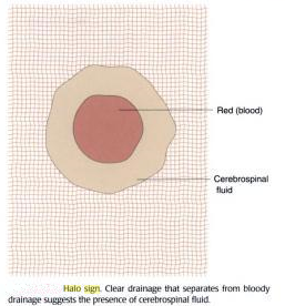
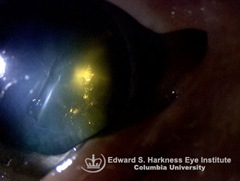
HALO SIGN/ HANDKERCHIEF SIGN - A finding in CSF rhinorrhea when CSF is mixed with Blood.

  • In patients with head trauma, a mixture of blood and CSF may make the diagnosis difficult.
  • CSF separates from blood when it is placed on filter paper, and it produces a clinically detectable sign: the ring sign, double-ring sign, or halo sign.
  • CSF will separate from blood when the mixture is placed on filter paper resulting in a central area of blood with an outer ring or halo.
  • Blood alone does not produce a ring.
  • The best ring is obtained with a 50: 50 mix of blood and CSF.
  • More importantly, they found that the presence of a ring was not exclusive for CSF.
  • Blood mixed with tap water, saline, and rhinorrhea fluid also produced a ring.
  • The halo sign does occur, but clearly does not clinch the diagnosis.

halo sign

HITSELBERGER’S SIGN - In Acoustic neuroma- loss of sensation in the postero-superior part of external auditory meatus supplied by Arnold’s nerve( branch of Vagus nerve to ear )

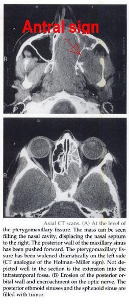
HOLMAN MILLER SIGN, ANTRAL SIGN-
The anterior bowing of the posterior wall of the antrum seen on lateral skull film .Pathognomic for juvenile nasopharyngeal angiofibroma.

antral sign

HONDOUSA SIGN–X-ray finding in Angiofibroma.
indicating infratemporal fossa involvement characterised by widening of gap between ramus of mandible and maxillary body.

HENNEBERT'S SIGN
It is a false positive fistula test when there is no evidence of middle ear disease causing fistula of horizontal semicircular canal. It is seen in 25% cases of  meniere's disease or congenital syphilis.In 25% cases of Meneire’s ,fibrous bands form connecting utricular macule to stapes footplate. In syphilis due to hypermobile stapes footplate.[Hennebert sign- pressure induced nystagmus, Hennebert symptom- pressure induced dizziness]

IRWIN MOORE’S SIGN——– positive squeeze test in chronic tonsillitis.

LAUGIER'S SIGN-Blood behind the eardrum suggests basilar skull fracture.

LEUDET'S SIGN-Inflammation of the eustachian tube can produce a bright clicking sound heard by the examiner through the otoscope while the patient experiences it as tinnitus.caused by reflex spasm of the tensor palati muscle.

LIGHT HOUSE SIGN—A small pin hole perforation with a pulsatile ear discharge is seen in Acute suppurative otitis media.

LYRE’S SIGN - splaying of carotid vessels( at junction of External & internal carotid artery) in carotid body tumor.

lyre sign

MILIAN’S EAR SIGN- Erysipelas can spread to pinna(cuticular affection), where as cellulitis cannot.

Cellulitis and erysipelas manifest as areas of skin erythema, edema and warmth in the absence of underlying suppurative foci.

They differ in that erysipelas involves the upper dermis and superficial lymphatics, whereas cellulitis involves the deeper dermis and subcutaneous fat.

As a result, erysipelas has more distinctive anatomic features than cellulitis; erysipelas lesions are raised above the level of surrounding skin, and there is a clear line of demarcation between involved and uninvolved tissue.

Classic descriptions of erysipelas note "butterfly" involvement of the face.

Involvement of the ear (Milian's ear sign) is a distinguishing feature for erysipelas since this region does not contain deeper dermis tissue.

erysipelas


OMEGA SIGN -INFANTILE OMEGA SHAPED EPIGLOTTIS SEEN IN LARINGOMALACIA.

omega epiglottis

Paul Dudley White's winking ear lobe sign-Movement of the ear lobe coincident with the pulse suggests tricuspid insufficiency.

PHELP’S SIGN - loss of crest of bone (as seen in CT-scan) between carotid canal and jugular canal in glomus jugulare.

RAT TAIL SIGN /“Bird-beak” sign-Sign in barium swallow of achalasia. The oesophagus is dilated, and contrast material passes slowly into the stomach as the sphincter opens intermittently. The distal oesophagus has a narrow segment and the image resembles a bird's beak.

rat tail

This is in contrast to the rat's tail appearance of carcinoma of oesophagus.Barium swallow shows characteristic rat tail appearance with irregular mucosa margins in carcinoma esophagus.

RISING SUN SIGN
There is red vascular hue seen behind the intact tympanic membrane. it is seen in glomus tumour, high jugular bulb and aberant carotid artery in the floor of middle ear.

RACCOON SIGN-Indicate subgaleal hemorrhage,and not necessarly base of skull .
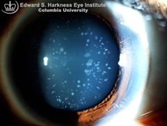
SCHWARTZ SIGN
It is also called flamingo flush sign. it is seen because of increased vascularity in submucous layer of promontory in active phase of otosclerosis(otospongiosis).

STEEPLE SIGN- X-ray finding in Acute laryngotracheobronchitis (CROUP).The steeple sign is produced by the presence of edema in the trachea, which results in elevation of the tracheal mucosa and loss of the normal shouldering (lateral convexities) of the air column. steeple

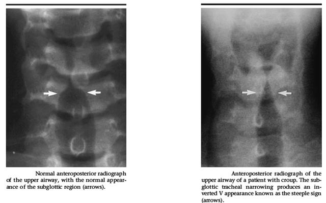
STANKIEWICK’S SIGN - indicate orbital injury during FESS. fat protrude in to nasal cavity on compression of eye ball from outside .

TEAR DROP SIGN

Seen in Orbital floor fracture. It is defined as tear drop shaped opacification seen hanging from the roof of the maxillary sinus on water's view. The floor of the orbit is the most common portion of the orbit to sustain fracture. A classic radiographic finding in blow-out fractures is the presence of a polypoid mass (the tear-drop) protruding from the floor of the orbit into the maxillary antrum The tear-drop represents the herniated orbital contents, periorbital fat and inferior rectus muscle.
orbit teardrop

THUMB SIGN
It is a thumb like impression (due to enlarged epiglottis) seen on X-ray lateral view neck in patients with acute epiglottitis.Direct visualization of the epiglottis by laryngoscope, if attempted, reveals a beefy red, edematous epiglottis.

thumb sign

TRAGUS SIGN
In acute otitis externa there is marked tenderness when tragus is pressed against the pinna.

TEA POT SIGN is seen in CSF rhinorrhoea.This could be related to the relationship of the sphenoid ostium to the sinus floor. The sphenoid ostium lies at an appreciable distance anterosupe-rior from the sinus floor. An increase in the CSF rhinorrhea therefore occurs in a case of sphenoid sinus leak when the patient bends forward as an increasing amount of CSF gains access to the ostium "teapot" sign.

teapot

uvula pointing sign - uvula points to side of palatal palsy

Uvula pointing sign- seen in rhinoscleroma .when scleroma involve nasopharynx ,uvula point towards roof of nasopharynx.

WOODS SIGN—– palpable jugulodigastric lymphnodes.


Bezold eponyms in ENT

  1. Bezold's triad
    1. Diminished perception of the deeper tones,
    2. retarded bone conduction, and
    3. negative Rinne's test,
    pointing, in the absence of objective signs, to otosclerosis
  2. Bezold's abscess
    abscess deep in the neck parapharyngeal space associated with suppuration in the mastoid cells
  3. Bezold's sign / symptom
    Inflammatory edema at the tip of the mastoid process in mastoiditis
  4. Bezold's ganglion
    An aggregation of nerve cells in the interatrial septum
  5. Bezold's mastoiditis
    Mastoiditis with perforation medially into the digastric groove and forming a deep neck abscess

Thursday, February 5, 2009

Heterochromia with miosis ?

heterochromia

Schistosomiasis

Schistosomiasis, also known as bilharziasis, results from long-lived infection by multicellular intravascular parasites of one of five trematode species — Schistosoma japonicum, S. mansoni, S. haematobium, S. intercalatum, or S. mekongi.

Parasite transmission and the consequent risk of human infection are strongly linked to specific geographic locations, because the parasite goes through several developmental stages that must occur in fresh water, including a period of growth within particular species of intermediate host snails.

  • Infection of humans with schistosoma species causes chronic hepatic and intestinal fibrosis or fibrosis, stricturing, and calcification of the urinary tract.
  • Infection follows contact with fresh water harboring larval parasites called cercariae, which penetrate humans' skin, become schistosomula, and enter capillaries and lymphatic vessels.
  • The worms migrate to the portal venous system, where they mature and unite, and then to the superior mesenteric veins, the inferior mesenteric and superior hemorrhoidal veins, or the vesical plexus and veins draining the ureters.
  • Eggs are produced and pass into adjacent tissues; many are shed in feces or urine.
  • The eggs hatch, releasing miracidia that infect freshwater snails, which ultimately release cercariae.

schistomiasisCheck this interactive graphics of Schistosomiasis.

Monday, February 2, 2009

Guess whats wrong in this pic?

4th nerve apsy

Answer: Three of the four children exhibit a left head tilt consistent with congenital 4th cranial nerve palsy. Right congenital fourth nerve palsy in all children except the one on the upper-right.


Quick Review: Congenital Fourth Nerve Palsies

  • Head tilt opposite the side of the palsy.
  • Facial asymmetry.
  • Large vertical fusional amplitudes.