Pages

Showing posts with label clinical image. Show all posts

Wednesday, July 1, 2009

Clinical Image – What are they?

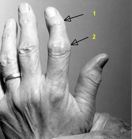
 heberden &bouchard nodes

1.Heberden’s nodes :

  • Heberden’s nodes are a clinical sign typically associated with osteoarthritis
  • Heberden’s nodes are often described as enlargements of the terminal (or distal) interphalangeal joints of the fingers
  • The histologic nature of the nodes is unknown.
  • They normally do not arise during the acute stage of osteoarthritic development, but rather during the chronic stage.In the acute stage, joints become erythematous, warm, and very tender to the touch. At this point, the joints are usually slightly swollen and may exhibit effusion. In addition, during the acute stage, pain in the area of the joints is often severe, occurring in spasms and with the sensation of burning and tingling in the skin overlying the joints. After several months, once the individual is in the chronic stage, the signs of inflammation disappear and bony outgrowths (or Heberden’s nodes) begin to arise over the terminal interphalangeal joints of the fingers. At this time, the joints are generally painless, and the characteristic deformation associated with Heberden’s nodes becomes obvious.

2.Bouchard's nodes :

  • Bouchard's nodes are comparable in presentation to Heberden's nodes, but are significantly less common.
  • They are hard, bony outgrowths on the proximal interphalangeal joints (the middle joints of fingers or toes.)
  • They are also a sign of osteoarthritis, and are caused by formation of bony outgrowths .

Thursday, May 21, 2009

Image challenge from NEJM

image

What is the most likely diagnosis?

1. Amyloidosis

2. Craniopharyngioma

3. Leukemia

4. Neuroblastoma

5. von Willebrand's disease


ANSWER:

4. Neuroblastoma

The image shows bilateral periorbital ecchymosis with dysconjugate gaze; the sclera appear normal. The "raccoon eyes" appearance is characteristically associated with neuroblastoma metastases to the skull. The appearance is not consistent with the other choices.


Check this previous post for further information – Raccoon eyes.

Monday, February 2, 2009

Guess whats wrong in this pic?

4th nerve apsy

Answer: Three of the four children exhibit a left head tilt consistent with congenital 4th cranial nerve palsy. Right congenital fourth nerve palsy in all children except the one on the upper-right.


Quick Review: Congenital Fourth Nerve Palsies

  • Head tilt opposite the side of the palsy.
  • Facial asymmetry.
  • Large vertical fusional amplitudes.

Sunday, January 4, 2009

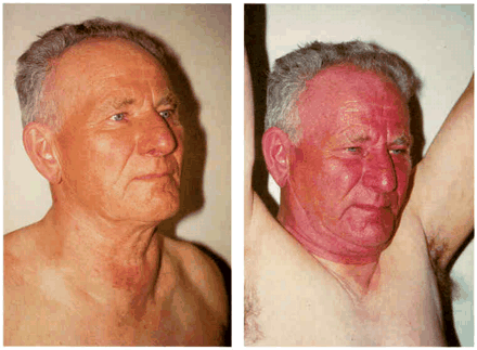
What is this ?The Pemberton Sign

Head and neck with arms down (left) and arms elevated (right).

pemberton sign


This is Pemberton sign positive.

  • With years of continued growth, the thyroid may extend downward and
  • enlarge within the chest, resulting in a substernal goiter.

  • Symptoms and signs may arise from compression of the structures located within the bony confines of the thoracic inlet, including the trachea, esophagus, and vasculature .
  • The Pemberton maneuver is a physical examination method that elicits manifestations of latent increased pressure in the thoracic inlet by altering arm position to further narrow the aperture.
  • The maneuver involves "elevating both arms until they touch the sides of the head"; if the sign is present, "after a minute or so, congestion of the face, some cyanosis, and lastly distress become apparent".
  • The Pemberton sign is an important indicator of increased pressure in the thoracic inlet.
  • Pemberton sign occurs when the thoracic inlet becomes obstructed during positional changes, resulting in compression of the jugular veins.

It is a common manifestation of retrosternal goiter but may also occur with lung carcinoma, lymphoma, thymoma, or aortic aneurysms.

Saturday, January 3, 2009

What is the diagnosis?

babesiosis

  • The peripheral-blood smear shows numerous intracellular organisms in red blood cells.
  • Multiple ring forms are seen, as well as rare tetrads (thin arrow).
  • These so-called Maltese cross formations are essentially pathognomonic of babesiosis.
  • The dark, round body in the right lower quadrant of the red blood cell with the tetrad is a Howell–Jolly body (thick arrow), an erythrocyte inclusion representing an incompletely extruded nucleus.
  • Howell–Jolly bodies are seen in patients with functional asplenia, and such patients are particularly susceptible to serious babesial and encapsulated bacterial infections.

Babesiosis is a worldwide tick-borne hemolytic disease that is caused by intraerythrocytic protozoan parasites of the genus Babesia.

The organisms are intraerythrocytic ring forms closely resembling Plasmodium, the organism causing malaria.

Three distinguishing features differentiate the two organisms.

    1. Babesial organisms usually form tetrads ("Maltese cross"),
    2. Do not have hemozoin pigments within the affected red blood cells and
    3. Have extracellular merozoites.

Babesia_LifeCycle

  • Atovaquone suspension (Mepron; 750 mg twice daily) plus azithromycin (Zithromax; 500 to 1,000 mg per day) is a very effective treatment.

Saturday, October 25, 2008

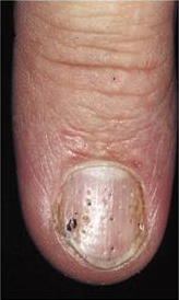
what is the diagnosis?

psoriasis


ans:Psoriatic nail disease

  • Nail involvement is common in psoriatic patients, affecting up to 80% of patients at some point in their lives
  • Nail psoriasis is common in psoriatic patients, particularly in patients with joint involvement.

The clinical findings associated with psoriatic nail disease correlate with the anatomical location of the nail unit that is affected by the disease.

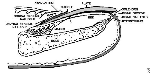
The nail unit is composed of the nail plate, the nail bed, the hyponychium, the nail matrix, the nail folds, the cuticle, the anchoring portion of the nail bed, and the distal phalangeal bones. nail

  • The nail plate is the largest component of the nail unit.
  • The nail matrix gives rise to the nail plate.
  • Any defect to the matrix results in onychodystrophy of the growing nail plate.
  • The proximal nail matrix forms the dorsal portion of the nail plate, whereas the distal matrix forms the ventral part of the nail plate.

The clinical presentation may vary depending on the location and the severity of inflammation of the affected nail unit.

Nail Signs and Anatomic Site of Disease

  • Nail matrix

Proximal: pitting, dystrophy:

  • Pitting is the most common manifestation in fingernails.originate from focal psoriasis of proximal matrix.Pitting is a result of the loss of parakeratotic cells from the surface of the nail plate.when focal psoriasis may become more marked then a pit may enlarge & produce a hole in nail plate ,a sign termed ELKONYXIS.
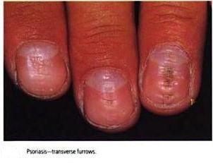
  • Beau lines: These lines are transverse lines in the nails due to books_002m, intermittent inflammation causing growth arrest lines
Distal:

  • leukonychia: Leukonychia is areas of white nail plate due to foci of parakeratosis within the body of the nail plate.
  • Nail bed
    • Onycholysis: Onycholysis is a white area of the nail plate due to a functional separation of the nail plate from its underlying attachment to the nail bed. It usually starts distally and progresses proximally, causing a traumatic uplifting of the distal nail plate. Secondary microbial colonization may occur.
    • Subungual hyperkeratosis: It is manifested as accumulated squames.in toenails subungual hyperkeratosis is the commonest finding.
    • Nail plate crumbling: Nail plate weakening due to disease of the underlying structures causes this condition.
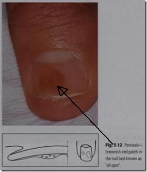
    • Oil drop or salmon patch/nail bed: This lesion is a translucent, yellow-red books_004b v discoloration in the nail bed resembling a drop of oil beneath the nail plate. This patch is the most diagnostic sign of nail psoriasis.
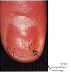
    • Splinter hemorrhage:Splinter hemorrhage/dilated tortuous capillaries in the dermal papillae. Splinter hemorrhages are longitudinal black lines due to books_00213minute foci of capillary hemorrhage between the nail bed and the nail plate. This is analogous to the Auspitz sign of cutaneous psoriasis, which is the pinpoint bleeding seen beneath the psoriatic plaques.
  • Nail fold

Paronychia


Tuesday, October 7, 2008

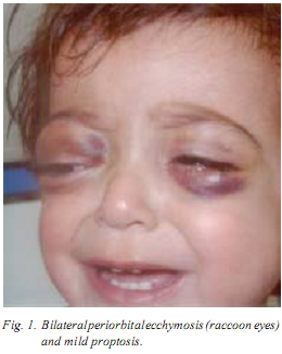
Clinical images-Raccoon eyes

raccoon eye


Raccoon eyes

A I5-month-old girl was admitted to hospital with the complaints of bruising around the eyes for 10 days. She had one month history of abdominal pain and accompanied by fever and vomiting for one week. Physical examination demonstrated pallor, periorbital ecchymoses (raccoon eyes) and bilaterally mild proptosis (Fig. 1). She also had a left sided abdominal mass, which was 5 × 8 cm in diameter. The laboratory investigation revealed hemoglobin level 6g/dL, white cell count 10,000/mm3 and platelet count 34,000/mm3 . Urinary vanillymandelic acid level was high. Bilateral bone marrow aspiration revealed infiltration with neuroblasts. Abdominal CT showed a left supra-renal mass (5 × 7 cm in diameter), which was diagnosed as a neuroblastoma on histo-pathologic examination.

  • The metastatic involvement of the periorbital tissues. has been described and the resultant proptosis and orbital ecchymosis has been given the tag of raccoon eyes’.
  • Orbital metastases can be found in up to 20% of children with stage IV neuroblastoma.
  • The characteristic "raccoon eyes" appearance associated with neuroblastoma and metastasis to the skull is probably related to obstruction of the palpebral vessels (branches of the ophthalmic and facial vessels) by tumor tissue in and around the orbits .

differential diagnosis:The correct diagnosis of this condition is sometimes delayed because of workup for child abuse or trauma. There are a multitude of differential diagnoses for the presentation of periorbital edema and ecchymosis,

 e.g.,

  1. child abuse or trauma,
  2. infection of the soft tissues associated with a spreading dental infection and an allergic reaction.
  3. Other systemic causes to be considered include myxoedema,
  4. other neoplasias such as lymphoma or
  5. haematological coagulopathies such as haemophilia.

note:

  • Neuroblastoma has been called the great mimicker because of its myriad clinical presentations related to the site of the primary tumor, metastatic disease, and its metabolic tumor by-products.
  • Pepper syndrome occurs in infants with overwhelming metastatic neuroblastoma of the liver that results in respiratory compromise. Pepper syndrome was identified as a localized primary tumor and metastatic disease limited to the skin, liver, and bone marrow in infants. Pepper syndrome has since been associated with stage 4S neuroblastoma, a unique entity that occurs only in infants younger than 1 year. Pepper syndrome generally confers a better prognosis, as it is associated with spontaneous regression.
  • "Blueberry muffin" babies are infants in whom neuroblastoma has metastasized to random subcutaneous sites. When provoked, the nodules become intensely red and subsequently blanch for several minutes thereafter. The response is probably secondary to the release of vasoconstrictive metabolic tumor by-products. These nodules can be diagnostic of neuroblastoma, but leukemic infiltrates that metastasize to the skin should be considered in the differential diagnoses when these children are evaluated.
  • Widespread metastasis of neuroblastoma to the bone may result in Hutchinson syndrome, which results in bone pain with consequent limping and pathologic fractures. Neuroblastomas that arise in the paraspinal ganglia may invade through the neural foramina, compress the spinal cord, and subsequently cause paralysis
  • The most common finding upon physical examination is a nontender, firm, irregular abdominal mass that crosses the midline. In contrast, children who present with Wilms tumor have a smooth mobile flank mass that typically does not cross the midline.