Pages

Showing posts with label medical high yield facts. Show all posts

Thursday, July 9, 2009

Eponyms for Percivall Pott


Eponym Description
Pott's aneurysm Arteriovenous aneurysm in which blood flows from an artery directly into a vein without going through a connecting sac
Pott's cancer Scrotal cancer originally described in chimney sweeps exposed to coal dust
Pott's disease Caries or osteitis of the vertebrae, usually of tuberculous origin (mycobacterium tuberculosis), characterized by softening and collapse of the vertebrae, often resulting in kyphosis, a hunchback deformity (Pott's curvature).
Pott's fracture Supramalleolar fracture of 1 or both ankle bones
Pott's gangrene Gangrene of foot from arterial insufficiency
Pott's paraplegia Paraplegia caused by spinal cord compression and abscesses in tuberculous spondylitis (Pott disease)
Pott's puffy tumor Pott puffy tumor is a subperiosteal abscess of the frontal bone that appears as a localized swelling of the overlying region of the forehead. It is the result of frontal sinusitis and osteomyelitis that erode through the anterior mantle of the frontal bone.

Wednesday, July 8, 2009

Splitting of Second heart sound

To understand Splitting of S2 better, we should first understand its normal physiology.

Second heart sound:

It has two audible components, the aortic valve closure sound (A2) and the pulmonic valve closure sound (P2), which are normally split on inspiration and virtually single on expiration.

Technique:

It has two audible components, the aortic closure sound (A2) and the pulmonic closure sound (P2), which must be separated by more than 20 msec (0.20 sec) in order to be differentiated and heard as two distinct sounds. It is clinically very important to determine the presence and degree of respiratory splitting and the relative intensities of A2 and P2.

Splitting is best identified in the second or third left ICS, since the softer P2 normally is confined to that area, whereas the louder A2 is heard over the entire precordium, including the apex.

Physiology:

Various research findings suggests that closure of the aortic and pulmonic valves initiates the series of events that produces the second heart sound. The main audible components, however, result from vibrations of the cardiac structures after valve closure. Using high-fidelity, catheter-tipped micro-manometers and echophonocardiography, it has been shown that the aortic and pulmonic valves close silently and that co-aptation of the aortic valve cusps precedes the onset of the second sound by a few milliseconds. The second sound therefore originates from after-vibrations in the cusps and in the walls and blood columns of the great vessels and their respective ventricles. The energy from these oscillations comes from sudden deceleration of retrograde flow of the column of blood in the aorta and pulmonary artery when the elastic limits of the tensed valve leaflets are met. This abrupt deceleration sets the whole cardiohemic system into vibration.

In order to understand splitting of the second heart sound, knowledge of its relationship to the cardiac cycle is essential.

First lets understand why is A2 before P2 ?

1.Both right & left ventricular systole ends at the same time .(ie, systolic muscle contraction of both ventricles ends at same time)

2.pulmonary arterial pressure is less than aortic pressure (i.e, pulmonary resistance to forward flow from ventricles is less than aortic resistance => therefore we can say that pulmonary impedance is less than aortic impedance.){impedance is nothing but resistance}

3.Therefore as pulmonary impedance is less, even after right ventricular systolic contraction blood continues to flow through valve until pulmonary arterial pressure increases more than right ventricle). But as aortic impedance is more ,it stops blood flow through the aortic valve before itself.

4.Due to the above reasons ,Right ventricular ejection begins prior to left ventricular ejection, has a slightly longer duration, and terminates after left ventricular ejection, resulting in P2 normally occurring after A2.

A2 and P2 are coincident with the incisura of the aorta and pulmonary artery pressure curves, respectively, and terminate left and right ventricular ejection periods.(incisura reflects closure of valves)

The differences between the aortic and pulmonary artery vascular impedance characteristics are also essential to understanding the effects of respiration on splitting of S2. When the pressure curves of the pulmonary artery and right ventricle are recorded simultaneously, the pulmonary artery curve at the level of the incisura (dicrotic notch) lags behind the right ventricular curve, or "hangs out" after it.

hangout interval

The duration of the "hangout interval" is a measure of impedance in the pulmonary artery system. In the highly compliant (low-resistance, high-capacitance) pulmonary vascular bed, the hangout interval may vary from 30 to 120 msec, contributing significantly to the duration of right ventricular ejection.

In the left side of the heart, because impedance is much greater, the hangout interval between the aorta and left ventricular pressure curves is negligible (less than or equal to 5 msec).

The hangout interval therefore correlates closely with impedance of the vascular bed into which blood is being injected. Its duration appears to be inversely related to vascular impedance.

Normal physiological splitting during respiration:

Alterations in the impedance characteristics of the pulmonary vascular bed and the right-sided hangout interval are responsible for many of the observed changes in splitting of S2.

In a normal physiologic setting, inspiration lowers impedance in the pulmonary circuit, prolongs the hangout interval and delays pulmonic valve closure, resulting in audible splitting of A2 and P2.

On expiration, the reverse occurs: pulmonic valve closure is earlier, and the A2–P2 interval is separated by less than 30 msec and may sound single to the ear. Since the pulmonary circulation has a much lower impedance than the systemic circulation, flow through the pulmonic valve takes longer than flow through the aortic valve. The inspiratory split widens mainly because of delay in the pulmonic component.

Traditionally it was believed that an inspiratory drop in intrathoracic pressure favored greater venous return to the right ventricle, pooling of blood in the lungs, and decreased return to the left ventricle. The increase in right ventricular volume prolonged right-sided ejection time and delayed P2; the decrease in left ventricular volume reduced left-sided ejection time and caused A2 to occur earlier. The delayed P2 and early A2 associated with inspiration, however, are best understood as an interplay between changes in the pulmonary vascular impedance and changes in systemic and pulmonary venous return. The net effect is that right ventricular ejection is prolonged, left ventricular ejection is shortened, and the A2–P2 interval widens during inspiration.

Clinical Significance

Normally the aortic closure sound (A2) occurs prior to the pulmonic closure sound (P2), and the interval between the two (splitting) widens on inspiration and narrows on expiration. With quiet respiration, A2 will normally precede P2 by 0.02 to 0.08 second (mean, 0.03 to 0.04 sec) with inspiration. In younger subjects inspiratory splitting averages 0.04 to 0.05 second during quiet respiration. With expiration, A2 and P2 may be superimposed and are rarely split as much as 0.04 second. If the second sound is split by greater than 0.04 second on expiration, it is usually abnormal.

Therefore, the presence of audible splitting during expiration (i.e., the ability to hear two distinct sounds during expiration) is of greater significance at the bedside in identifying underlying cardiac pathology than is the absolute inspiratory increase in the A2–P2 interval.

ABNORMAL SPLITTING OF THE SECOND HEART:

(1) persistently single;

(2) persistent (audible expiratory) splitting, with normal respiratory variation;

(3) persistent splitting without respiratory variation (fixed splitting); and

(4) reversed (paradoxical) splitting.

splitting s2

(1) persistently single;

  • When S2 remains single throughout the respiratory cycle, one component is absent or the two components are persistently synchronous.
  • The most common cause of a single S2 is inaudibility of the P2 in older adults with increased anteroposterior chest dimensions.
  • In the setting of
    congenital heart disease, a single S2 due to absence of the pulmonary component is a feature of pulmonary atresia, severe pulmonary valve stenosis, dysplastic pulmonary valve, or complete transposition of the great arteries.
  • Conversely, a single S2 due to inaudibility of the A2 occurs when the aortic valve is immobile (severe calcific aortic stenosis) or atretic (aortic atresia).

(2) persistent (audible expiratory) splitting, with normal respiratory variation;

  • Persistent splitting may be due to a delay in P2, as in cases of simple complete right bundle branch block,or to early timing of the A2, as occasionally occurs in cases of mitral regurgitation(since early emptying of left ventricle –> early closure of Aortic valve).
  • Normal directional changes in the interval of the split (greater with inspiration, lesser with exhalation) in the presence of persistent audibility of both components defines the split as persistent but not fixed.
  • these conditions have wide inspiratory split

(3) fixed splitting;

  • This term applies when the interval between the A2 and P2 is not only wide and persistent but also remains unchanged during the respiratory cycle.
    Fixed splitting is an auscultatory hallmark of  atrial septal defect(ASD). 
  • A2 and P2 are widely separated during exhalation and exhibit little or no change in the degree of splitting during inspiration.

Reason behind wide splitting:

The wide splitting is caused by a delay in the P2 because a marked decrease in pulmonary vascular impedance prolongs the interval between the descending limbs of the pulmonary arterial and right ventricular pressure pulses (“hangout”), and therefore delays the pulmonary incisura and the P2.

Reason behind fixed splitting:

  • We know that in normal individuals , the amount of pulmonary ejection is more during inspiration compared to that in expiration (since pulmoary impedance decreases during inspiration)
  • In ASD .the amount of blood which passes throught he pulmonary valve ramains the samee in both inspiration& expiration because of Phasic changes in systemic venous return during respiration in patients with atrial septal defect are associated with reciprocal changes in the volume of the left-to-right shunt, minimizing respiratory variations in right ventricular filling. (ie, whenever venous return increases in inspiration, it causes a reciprocal decrease in left to right shunting in atria & whenever venous return decreased as in expiration ,shunting increases =>this maintains equal amount of blood in right ventricle irrespective of inspiration or exoiration)
  • The net effect is the characteristic wide, fixed splitting of the two components of the S2.

(4) reversed (paradoxical) splitting. 

  • This term refers to a reversed sequence of semilunar valve closure, the P2 preceding the A2.
  • Common causes of paradoxical splitting are complete left bundle branch block or a right ventricular pacemaker, both of which are associated with initial activation of the right side of the ventricular septum, and delayed activation of the left ventricle owing to transseptal (right-to-left) depolarization.
  • When the S2 splits paradoxically, its two components separate during exhalation and become
    single (synchronous) during inspiration .
  • Inspiratory synchrony is achieved as the two components fuse because of a delay in the P2, less to earlier timing of the aortic
    component.

  splitting s2


Please review this article & if you have any doubts do tell me.

Sunday, June 28, 2009

Eponymous Signs of Aortic Regurgitation

Austin Flint Murmur

Description

The murmur typically begins in mid-diastole, often has a presystolic accentuation, and terminates at the end of diastole. It is low-pitched, with a rough and rumbling quality, and best heard at the apex. An Austin Flint murmur can be deemed present only in the setting of aortic regurgitation without coexisting mitral stenosis, since the latter can generate a similar murmur.

Pathophysiology

Austin Flint postulated that regurgitant blood flow in severe aortic regurgitation impinges on the leaflets of the mitral valve, leading to a functional stenosis. Diastolic inflow across this narrowed mitral valve orifice generates turbulence that is clinically appreciable as a mid- to late diastolic murmur . Later investigations have advanced a variety of other theories as causes for the murmur, including overlap of aortic regurgitation and mitral inflow jets , fluttering of mitral valve leaflets (, and left ventricular endocardial vibrations due to the aortic regurgitation jet. However, a universally accepted explanation remains elusive.

Elicitation

The murmur is best heard on auscultation at the apex by using the bell of the stethoscope, with the patient in the left lateral position.


Corrigan Pulse:

Description

Large volume collapsing pulse of the carotid artery

Pathophysiology

A recent investigation found that patients with aortic regurgitation had increased amplitude of the pulse, lower mean arterial pressure, and narrower pulse pressure than normal patients . The investigators concluded that these characteristics reflected an increase in the compliance of the arterial wall in patients with aortic regurgitation.

Elicitation

The examiner palpates the patient's radial artery while elevating the wrist. If the pulse clearly increases in amplitude, then the sign is present.


Duroziez Sign

Description

The sign denotes an intermittent to-and-fro femoral artery murmur (occurring in systole and diastole, respectively) generated by femoral artery compression.

Pathophysiology

Duroziez believed the systolic portion of the murmur was caused by forward flow into the lower extremity and that the diastolic segment was caused by aortic regurgitation toward the heart.

Elicitation

Duroziez auscultated the femoral artery while applying digital compression proximal and distal to the stethoscope .Blumgart and Ernstene  ) replaced digital compression with cephalad and caudad tilting of the stethoscope.


Hill Sign:This sign is also known as the popliteal–brachial gradient.

Description

A 20 mmHg difference in the popliteal and brachial systolic cuff pressures

Pathophysiology

The Hill sign, therefore, remains an unexplained artifact of indirect blood pressure measurement that is consistently more common and pronounced in patients with aortic regurgitation than in those without.

Elicitation

The blood pressure is manually obtained over the brachial and femoral arteries by using appropriately sized cuffs, with the patient in the recumbent position. The difference in systolic pressures denotes the gradient.


Watson’s water hammer pulse: also known as collapsing pulse, cannonball pulse or pulsus celer.

Description

Large-volume, ‘collapsing’ bounding peripheral pulses.

Pathogenesis        

The abrupt jerky, forceful upstroke of the whp implies a rapid filling of the radial artery in systole due to an extra large amount of blood pushed by the distended left ventricle into relatively empty arterial vessels. The collapsing or sudden down stroke may be partly due to a sudden fall in the diastolic pressure in the aorta due to regurgitation of blood into the left ventricle through a leaky valve and partly due to the rapid emptying of the arterial system due to the marked increase in the velocity of the bloodstream. Apart from the pathogenetic factors which cause the widened pulse pressure, lifting the patient's arm vertically upwards when eliciting the whp, helps the blood to empty quickly into the heart during diastole due to the gravity and also brings the radial artery more in line with the outflow stream of the aorta, thus accentuating the sign.


Traube’s sign/pistol shot sounds :

Booming systolic and diastolic sounds heard over the femoral artery when it is compressed distally

De Musset’s sign: Head nodding with each heart beat

Quincke’s sign :Pulsations in the nail capillary bed seen when light is transmitted through the fingertips or exerting gentle pressure on the tip of a fingernail .

Lighthouse sign:Blanching and flushing of forehead

Landolfi’s sign :Alternating constriction and dilatation of pupil

Becker’s sign :Visible pulsations of the retinal arterioles

Mueller’s sign :Visible pulsations of uvula

Mayen’s sign :Diastolic drop of BP >15 mmHg with arm raised

Rosenbach’s sign :Pulsatile liver

Lincoln sign :Pulsatile popliteal artery

Gerhardt’s sign : Pulsatile spleen

Sherman sign: Prominently located and palpated dorsalis pedis pulse

Monday, June 22, 2009

Important Murmurs

Austin Flint murmur : Austin Flint murmur is a mid-diastolic rumbling audible in subjects with severe aortic regurgitation that is best heard at the apex with little radiation. Several theories have been suggested for the origin of the murmur: (1) vibration of the anterior mitral valve leaflet due to the regurgitant jet, (2) collision of the jet with mitral inflow, (3) increased mitral inflow velocity due to narrowing of the valve orifice by the jet, and (4) vibration from the jet impinging on the myocardial wall. It differs from mitral stenosis murmur in that ,it occurs in the presence of a murmur of aortic valve insufficiency and in the absence of the rheumatic, mitral opening snap.

Carey Coombs murmur : Mitral valvulitis associated with acute rheumatic fever may cause a low-pitched mid-diastolic rumble. It can be differentiated from the diastolic murmur of Mitral stenosis by the absence of (1)an opening snap, (2)presystolic accentuation & (3)loud first heart sound.

Cruveilhier-Baumgarten murmur : Venous hum heard in epigastric region (on examination by stethoscope) due to collateral connections between portal system and the remnant of the umbilical vein in portal hypertension.

Duroziez's murmur : Its a to & fro murmur heard over the femoral artery during both systole& diastole. It is elicited by applying gradual arterial compression with the diaphragm of the steth. This compression not only produces systolic murmur(which is the normal result of arterial compression) but also a diastolic murmur(which is pathologic & suggestive of aortic regurgitation).Sensitivity of 58-100%.False positives occur in high output states. In high output states the double murmur is due to forward flow. In Ar one murmur is due to forward flow& the other due to reverse flow.The two can be differnetiated by applying pressure first on the more cephalad edge of the diaphragm & then on its more caudal edge. The murmur of forward flow is enhanced by compressing the cephalad edge. Conversely the reverese flow murmur is enhanced by compressing the caudad edge.

Gibson murmur :The typical continuous "machinery-like" murmur of patent ductus arteriosus.train in tunnel murmur

Graham Steell's murmur : Due to pulmonary regurgitation in patients with pulmonary hypertension and mitral stenosis. It is a high pitched early diastolic murmur heard best at the left sternal edge in the second intercostal space with the patient in full inspiration.

The murmur is heard due to a high velocity regurgitant flow across the pulmonary valve; this is usually a consequence of pulmonary hypertension. The Graham Steell murmur is often heard in patients with chronic cor pulmonale as a result of chronic obstructive pulmonary disease.

Means-Lerman "scratch" murmur: Increased flow across the pulmonary valve in Thyrotoxicosis may be associated with ejection systolic murmur.The ejection systolic murmur owing to hyperthyroidism may have a scratchy quality (Means-Lerman scratch), and, frequently, the intensity of P2 is increased because of mild to moderate pulmonary hypertension.

Roger's Murmur:A loud pansystolic murmur caused by interventricular septal defect of the heart; maximal at the left sternal border.

Seagull murmur  a raucous murmur with musical qualities, such as that heard occasionally in aortic insufficiency.A "seagull’s cry murmur" is defined as a murmur imitating the cooing sound of a seagull. This type of murmur is typically characterized by a musical timbre and a high frequency, and may occur as a result of various valve diseases. It is usually described as a sign of tight calcific aortic stenosis, when the murmur’s high frequency components are transmitted to the lower left sternal border and the cardiac apex during most of systole (Gallavardin’s phenomenon). In this condition, the typical harsh timbre of the ejective murmur tends to assume a musical high pitched quality, resembling that of mitral regurgitation, which may be reminiscent of the cry of a seagull. A protodiastolic murmur with similar characteristics, typically in decrescendo, may occur in severe aortic valve regurgitation, particularly when the regurgitant flow presents high velocities. However, a seagull’s cry murmur may also be the sign of mitral regurgitation or prolapse. Similarly, the musical and holosystolic sound reflects the presence of high frequency components due to high velocities of reflow.
Still's murmur An innocent musical murmur resembling the noise produced by a twanging string; almost exclusively in young children, of uncertain origin and ultimately disappearing.{There are five innocent murmurs of infancy and childhood: (i) pulmonary flow murmur, (ii) Still's murmur, (iii) venous hum, (iv) carotid bruit, (v) physiologic pulmonary branch stenosis murmur of neonate}

Wednesday, June 17, 2009

Wednesday, June 10, 2009

Babinski sign- Mechanism& other Babinski like responses.

The Babinski sign
This eponym refers to the dorsiflexion of the great toe with or without fanning of the other toes and withdrawal of the leg, on plantar stimulation in patients with pyramidal tract dysfunction. ni_2000_48_4_314_1509_2

The characteristic response is dorsiflexion of the great toe by recruitment of extensor hallucis longus (EHL) muscle.

The art of elicitation:
The reflexogenic area for the plantar reflex is the first sacral (S1) dermatome with the receptor nerve endings being located in the skin. The afferent nerve is the tibial nerve, the spinal cord segments involved in the reflex arc being 4th and 5th lumbar and 1st and 2nd sacral.

Position
All the leg muscles should be visible and in a relaxed state. This can be achieved by positioning the patient in a way that the knee is slightly flexed and the thigh is externally rotated. The patient should be warned that the sole is going to be scratched and ask him to try to let his limb remain as floppy as possible. The toes should not be touched at all.

Stimulation
Any part of the leg can be stimulated, but the best technique is to stimulate the lateral plantar surface and the transverse arch in a single movement upto the middle metatarsophalangeal joint with a firm applicator lasting 5 to 6 seconds. Difficulties are bound to arise in certain clinical situations which makes elicitation and interpretation of plantar response inconclusive. It is imperative that one realises these problems and be aware of their solutions in order to arrive at an appropriate clinical conclusion [Table II].

ni_2000_48_4_314_1509_1

Interpretation of the response
The plantar response may be:
1. Normal flexor plantar response
2. Pathologic or abnormal extensor plantar response (Babinski’s sign)


Normal flexor plantar response
In normal people after infancy, there is a plantar flexion of the foot and toes along with adduction of the toes. The primary movement is a plantar flexion of the great toe at the metatarsophalangeal joint, even if the terminal joint appears to extend. The response is a fairly rapid one and may be accompanied at times by an associated flexion of the hip and knee on the stimulated side.
Abnormal extensor plantar response (Babinski’s sign)
The Babinski’s sign is encountered in patients with pyramidal tract dysfunction and is characterised by a dorsiflexion or extension of the great toe with or without fanning or abduction of the other toes. The fully developed response is also accompanied by dorsiflexion of the ankle and flexion of the hip and knee joint and slight abduction of the thigh, leading to a withdrawal of the leg on plantar stimulation.
The Babinski sign is always pathological. There is no such thing as a negative Babinski sign.

mechanism:

In most mammals the limbs are automatically retracted on painful stimulation as a defence reflex, which is more pronounced in hind limbs. Sherrington called it, the flexion reflex synergy, because activation of all muscles effected shortening of the limb; the toe extensors forming part of this shortening synergy. Confusion has arisen from the application of the term extensor plantar response to a movement which forms part of a flexion synergy of the lower limbs. The toe 'extensors' although named extensors by anatomists, are infact flexors in a physiological sense because their action is to shorten the limb and contract reflexly along with other flexor muscles.

The Babinski sign may be a normal occurrence in the first year of life. In the infant, before  myelination of the nervous system is complete and an upright stance has been achieved, the normal plantar response is extensor, due to a brisker ‘flexion synergy’as part of the withdrawal response to pain. As the nervous system matures and the pyramidal tracts gain more control over spinal motor neurons, the ‘flexion synergy’ becomes less brisk and the toe ‘extensors’ are no longer a part of it. When the child assumes an upright posture, the plantar response becomes part of the postural reflex maintaining the tones of the foot and leg. At this time, the normal response to stimulation becomes a flexor movement of the toes and the ‘withdrawal extensor’ movement is suppressed by the influence of the pyramidal tract over the spinal reflex arc. The toe then goes down instead of up, as a result of a segmental reflex involving small foot muscles and the overlying skin.This is considered to be normal in adults and is termed - flexor plantar response. With lesions of the pyramidal system, structural or functional, this segmental downward response of the toes disappears, the flexion synergy may become disinhibited and the EHL muscle is again recruited into the flexion reflex of the leg producing the sign of 'Babinski'.
The pyramidal tract thus maintains a suppressor action on the ‘flexion reflex’synergy. Pyramidal tract dysfunction however, allows the response to revert to the withdrawal movement by releasing or facilitating the ‘flexion reflex synergy’ of which contraction of the extensor hallucis longus muscle forms an integral part. A Babinski sign can appear only if the intraspinal pathways of the ‘flexion reflex synergy’are operative, however severe the motor deficit in the foot. The motor neurons of the
leg muscles are laminated into separate columns within the anterior horns of the cord, each of which supply proximal or distal flexor or extensor muscles. Both structural as well as functional lesions of the pyramidal tract fibres projecting onto the lumbosacral anterior horn cells and interneurons supplying the leg muscles subserving the ‘flexion reflex synergy’ can release the
Babinski sign. Reversible pathophysiologic conditions result in, or produce, a transient extensor plantar response.Structural lesions produce more lasting effects

The muscles taking part in a fully developed response include extensor hallucis longus, tibialis anterior, extensor digitorum longus, hamstring group of muscles and tensor faciae latae. The characteristic response is dorsiflexion (extension) of the [big toe], which precedes all other movements. It is followed by fanning out and extension of the other toes, dorsiflexion of the ankle and flexion of the hip and knee joint. This response represents 'positive' Babinski sign. There is no such thing as a 'negative' Babinski sign.

The dorsiflexion of the toes may be the only visible effect, but the contraction of the thigh and leg muscles is always present and can be detected by palpation. Contraction of the tensor fasciae
latae has been referred to as Brissaud’s reflex.
So the fully developed extensor plantar response forms part of the primitive ‘flexion reflex synergy’ of the lower limbs designed to withdraw the limb from a painful stimulus.
This spinal defence reflex mechanism described by Sherrington, activates all the muscles involved in
shortening the stimulated limb. It involves flexion of the hip and knee, dorsiflexion of the ankle and extension of the great toe. The ‘toe and foot extensors’ although named extensors by anatomists, are in fact flexor in a physiological sense, because their action is to shorten the limb and
contract reflexly along with other flexor muscles. The physiologist looks on the Babinski sign as simply a part of the ‘primitive flexion reflex’.

The function of the pyramidal tract may not only be disturbed by structural lesions of myelin sheaths, axons, or both, but also by non-neurological conditions [Table I].ni_2000_48_4_314_1509_3

Types of Babinski sign
a) Minimal Babinski sign : Contraction of hamstring muscles and tensor faciae latae.

b) True Babinski sign : Includes all the components of the fully developed extensor plantar reflex.

c) Pseudo Babinski sign : One may encounter this type of response in sensitive individuals, plantar hyperaesthesia, and choreo-athetosis due to hyperkinesis. True Babinski can be clinically distinguished from the false Babinski by the contraction of hamstring muscles in the former, and failure to inhibit the extensor response by pressure over the base of the great toe. The true Babinski sign is reproducible, unlike voluntary withdrawal of the toes.

d) Exaggerated Babinski sign : It can either be in the form of 'flexor spasm' or 'extensor spasm', depending upon the muscles i.e. whether flexors or extensors, have excess of tone. Flexor spasms occur in spinal cord disease, bilateral upper motor neuron lesion at a supraspinal level, multiple sclerosis and subacute combined degeneration of the cord, while 'extensor spasm' occurs in patients with corticospinal tract lesion when the posterior column function is normal.

e) Inversion of plantar reflex : If the short flexors of the toe are paralysed or flexor tendons are severed accidentally, an extensor response may be obtained.

f) Tonic Babinski reflex : Characterised by slow prolonged contraction of extensors of toe, seen in frontal lobe lesions and extrapyramidal involvement.

g) Crossed extensor response/bilateral Babinski sign : Unilateral stimulation produces bilateral
Babinski in patients with bilateral cerebral disease and spinal cord disease.

h) Spontaneous Babinski : In infants and children following manipulation of the foot, and in patients with extensive pyramidal tract diseases, passive extension of the knee or passive flexion of the hip and the knee, may produce a positive Babinski sign.

Alternate methods
The late 19th and early 20th century was abound with disclaimers associated with founders of new reflex movements of the great toe. These movements are known by the term 'Babinski like responses'. These responses can be elicited by the following techniques, each with its own eponym.

  1. Chaddock's sign:The extension of the big toe may be obtained by stimulating the dorsal lateral aspect of the foot from the posterior portion of the skin beneath the external malleolis anteriorly along the external edge of the foot.
  2. Gordon's sign: squeezing the calf muscle
  3. Oppenheim sign: applying pressure along the shin of tibia
  4. Gonda's sign: pressing the 4th toe downwards and then releasing it with a snap
  5. Stransky sign: vigorous adduction of the little toe followed by its sudden release
  6. Schaefer's sign: squeezing the Achilles tendon
  7. Rossolimo's sign: flexion of the toes, on quick percussion of the tips of the patients toes with the finger tip
  8. Mendal Bechtrew sign: flexion of the four outer toes induced by tapping the dorsum of the foot in the region of cuboid bone
  9. Bing's sign:  giving multiple pinpricks on dorsolateral surface of the foot
  10. Moniz sign: forceful passive plantar flexion of the ankle
  11. Throckmortan sign: pressing over the dorsal aspect of the metatarsophalangeal joint of the great toe
  12. Strumpell sign: application of forceful pressure over anterior tibial region
  13. Cornell sign: scratching the dorsum of the foot along the inner side of the extensor tendon of the great toe
  14. plantar flexion and fanning of the toes on tapping the mid plantar region of the foot or base of the heel.

Most of these signs imply an increase in the reflexogenous zone and denote responses from different parts of the receptive field. When sufficiently facilitated, the reflex may be elicited by other stimuli as well. Infact, in extreme cases of UMN deficit, the complete ‘flexion reflex’ may be exhibited spontaneously and continuously; the patient lies in bed, the hip and knee flexed, and the ankle and great toe dorsiflexed. In other cases of severe UMN deficit, almost any unpleasant stimulus, such as scratching, pinching, or pricking, will evoke the ‘flexion reflex’, even when applied as high as the thigh, far from the usual reflexogenous zone.


Fallacies
An extensor response may be present when there is no damage to the pyramidal tract. A possible explanation being the excitation of the distal motor neurons and inhibition of the impulses via flexor reflex afferent
nerve fibres can be dissociated because they are mediated by different neurons, however closely linked. On the contrary, cases with proven damage to the pyramidal system have had normal plantar response. We should understand that corticospinal fibres not only originate in different parts of the cortex, but also have different terminations. Babinski sign can be expected only when 'leg fibres' of the pyramidal tract are involved. Plantar areflexia can be noted in cases with loss of sensation of sole due to lesion of the first sacral cutaneous distribution. The same can be observed in paralysis of extensors or long flexors of great toe. In spinal shock, cessation of tonic discharge of spinal neurons by excitatory impulses in descending pathways may explain its non existence. Drugs like parenteral physostigmine in physiological doses may also abolish a plantar response.

    »   Conclusion        

Despite the continuing controversy and observer bias, clinical utility of Babinski sign remains unchallenged. The role of pyramidal system in the pathophysiology of this sign is quite clear. Pyramidal tract dysfunction releases the flexion reflex synergy, of which contraction of the extensor hallucis longus muscle forms an integral part. The most important and vital question in interpreting the plantar response is not, whether the great toe goes up or not, but is whether an upgoing toe is pathological or not. For an appropriate answer the method of observation is much more important than the method of elicitation.


Modified From--Kumar SP, Ramasubramanian D. The Babinski sign--a reappraisal. Neurol India 2000;48:314

Thursday, June 4, 2009

Respiratory Sinus Arrhythmia- Why Does the Heartbeat Synchronize With Respiratory Rhythm?


Respiratory sinus arrhythmia (RSA) :

It is a naturally occurring variation in heart rate that occurs during a breathing cycle. Heart rate increases during inspiration and decreases during expiration.

It is one of the physiologic interactions between respiration and circulation, where heart rate variability in synchrony with respiration, by which the R-R interval on an ECG is shortened during inspiration and prolonged during expiration occurs.

A clustering of heartbeats (R waves of an ECG) during inspiration and a scattering during expiration are clearly seen.

CLINICAL SIGNIFICANCE:

studies have shown that the efficiency of pulmonary gas exchange is improved by RSA, suggesting that RSA may play an active physiologic role. The matched timing of alveolar ventilation and its perfusion with RSA within each respiratory cycle could save energy expenditure by suppressing unnecessary heartbeats during expiration and ineffective ventilation during the ebb of perfusion. Furthermore, evidence has accumulated of a possible dissociation between RSA and vagal control of that heart rate, suggesting differential controls between the respiratory modulation of cardiac vagal outflow and cardiac vagal tone. RSA or heart rate variability in synchrony with respiration is a biological phenomenon, which may have a positive influence on gas exchange at the level of the lung via efficient ventilation/perfusion matching.

respiratory sinus arrhythmia

Scheme showing the conceptual effects of RSA (top) and its inversion (bottom) on the relationship between alveolar gas volume and capillary blood flow during inspiration (left) and expiration (right). Curved horizontal arrows and vertical arrows indicate the volume of blood flow circulating in the pulmonary capillary bed and the direction of alveolar gas interfacing with the pulmonary capillary blood. V/Q = ventilation/perfusion.

Mechanisms of RSA :

Heart rate is determined by the firing frequency of the sinus node of a cardiac pacemaker. This frequency is determined by the balance between the cardiac sympathetic and vagal activities to the sinus node.

The activity of the cardiac vagal nerve is assumed to be modulated by respiration, and hence the sinus node activity is secondarily modulated by the respiratory rhythm. Regarding the genesis of RSA, both the respiratory and circulatory centers in the brainstem appear to be responsible. Moreover, projections from the cerebral cortex, limbic system, and other parts of the brain to the brainstem should exist.

In mammals, the following two major mechanisms have been recognized for generating RSA:

  1. direct modulation of the cardiac vagal preganglionic neurons by central respiratory drive;   The cardiac vagal efferent fibers are fired preferentially during expiration, and this respiratory-related activity is maintained even after the vagal nerve is resected at the peripheral to the recording site.The vagal efferent fibers are more powerfully excited during expiration by stimulating the arterial chemoreceptors and baroreceptors.Respiratory modulation could also be mediated by gating of the excitatory reflex inputs into the preganglionic neurons. Indeed, the membrane potential of cardiac vagal preganglionic neurons has been demonstrated to be hyperpolarized during each inspiration due to the arrival of an acetylcholine-mediated inhibitory postsynaptic potential, which makes neurons less amenable to excitatory inputs during inspiration.
  2. and inhibition of cardiac vagal efferent activity by lung inflation.On the other hand, afferent activity arising in the lungs is also an important mechanism with which to generate RSA.Lung inflation inhibits cardiac vagal efferent activity and evokes tachycardia by stimulating the pulmonary C-fiber afferents (ie, pulmonary stretch receptors). This effect may be so strong that it reverses the bradycardia evoked by arterial chemostimulation into a tachycardia. The efferent cardiac vagal nerve plays the major role in the genesis of RSA, whereas the contribution of the cardiac sympathetic nerve seems to be minimal. During inspiration, as described above, the activity of the efferent cardiac vagal nerve is almost abolished. Hence, the R-R interval on an ECG is shortened. In contrast, during expiration, the activity of the efferent cardiac vagal nerve reaches its maximum, thus extending the R-R interval. The difference in the R-R interval between inspiration and expiration can be regarded as an indication of the magnitude of RSA, which is assumed to reflect the cardiac vagal outflow within its physiologic range.Accordingly, the magnitude of RSA has been widely used as a clinical measure of cardiac vagal activity. A neural basis for RSA has been demonstrated by its elimination or substantial attenuation following cervical vagotomy, ganglionic blockade, cholinergic blockade, and heart transplantation.


Abridged from CHEST journal.

Wednesday, June 3, 2009

Laennec - eponyms

Laennec, a French physician and the inventor of the stethoscope.

Laennec's cirrhosis :Other name for Alcoholic Cirrhosis .Most common form of liver cirrhosis.refers to the appearance of regenerated liver, comprising small lobules separated by a fine, fibrous tissue.

Laennec's catarrh: a form of bronchial asthma characterized by the discharge of small, round, beadlike bodies of  mucus called Laennec's pearls.

Laennec's thrombus : it is an antenatal thrombus in the heart
Laennec's pearls : refers to beadlike mucus bodies produced by asthmatics.

Tuesday, April 14, 2009

Carpal tunnel syndrome – Important signs.

Phalen sign:Hyper flexion of the wrist for 60 seconds may elicit paresthesia in the median nerve distribution.

Tinel sign :Tapping the volar wrist over the median nerve may produce paresthesia in the median distribution of the hand.

Flick sign :Shaking or flicking one's hands for relief during maximal symptoms.

Friday, February 20, 2009

Filarial dance sign

A 35-year-old man presented with right-sided scrotal pain. Clinically, the right epididymis was thickened. High resolution ultrasound (10MHz) examination showed four cystic spaces in the right epididymal region and one in the left epididymal region measuring 2 mm to 8 mm with multiple small (<1mm) objects exhibiting peculiar random movements as described by the Filarial Dance Sign.

  • Lymphatic filariasis is a major health problem in India with most infections caused by Wuchereria bancrofti. The presence of adult worms of Wuchereria bancrofti in the infected individuals is confirmed by detecting microfilariae or filarial antigens in the patient's blood .
  • Ultrasound scans have been used to detect living adult W. bancrofti on account of the characteristic pattern of adult worm movements, known as the filarial dance sign in dilated intrascrotal juxtatesticular lymphatics (worm nests) of approximately 80% of microfilaremic but asymptomatic men residing in endemic areas.
  • Ultrasound is a valuable tool in the diagnosis of cases of lymphatic filariasis. Amaral et al  had first reported the use of ultrasound to visualize adult worms of W. bancrofti in the scrotal area of infected men. They described a continuous, distinctive and specific pattern of worm movement called the "Filarial dance" sign. In patients who exhibited this sign, nests of adult W. bancrofti were found in the lymphatic vessels of the spermatic cord on surgery.
  • The typical movement of these filariae, called the filarial dance sign (FDS), provides an opportunity to observe the adult worms in vivo.
  • The characteristic movements of adult filarial worms are called the filarial dance sign (FDS) and are a reliable diagnostic finding.

Also check my other detailed article on TPE( Tropical Pulmonary Eosinophilia)

Thursday, February 5, 2009

Schistosomiasis

Schistosomiasis, also known as bilharziasis, results from long-lived infection by multicellular intravascular parasites of one of five trematode species — Schistosoma japonicum, S. mansoni, S. haematobium, S. intercalatum, or S. mekongi.

Parasite transmission and the consequent risk of human infection are strongly linked to specific geographic locations, because the parasite goes through several developmental stages that must occur in fresh water, including a period of growth within particular species of intermediate host snails.

  • Infection of humans with schistosoma species causes chronic hepatic and intestinal fibrosis or fibrosis, stricturing, and calcification of the urinary tract.
  • Infection follows contact with fresh water harboring larval parasites called cercariae, which penetrate humans' skin, become schistosomula, and enter capillaries and lymphatic vessels.
  • The worms migrate to the portal venous system, where they mature and unite, and then to the superior mesenteric veins, the inferior mesenteric and superior hemorrhoidal veins, or the vesical plexus and veins draining the ureters.
  • Eggs are produced and pass into adjacent tissues; many are shed in feces or urine.
  • The eggs hatch, releasing miracidia that infect freshwater snails, which ultimately release cercariae.

schistomiasisCheck this interactive graphics of Schistosomiasis.

Wednesday, January 21, 2009

List of Medical Eponyms

512ZA3FPQ5L._SS500_

Medical eponyms are always favorite choice of Questions be it for viva-voice or in Mcqs in competitive exams .So be thorough with them ,in the file provided you will have A-Z list of all medical eponyms .So read them well.

Download MEDICAL EPONYMS pdf file from 4Shared.


Check my other post for Eponymous signs in ENT.

Tuesday, November 4, 2008

Picibanil

A lyophilized preparation of a low-virulence strain (SU) of Streptococcus pyogenes (S. hemolyticus), inactivated by heating with penicillin G. It has been proposed as a noncytotoxic antineoplastic agent because of its immune system-stimulating activity.

Monday, October 13, 2008

Bacterial Meningitis-high yield facts

  • The classic triad of symptoms of bacterial meningitis includes
    •  fever,
    • stiff neck,
    • and alterations of mental status.
  • However, only two thirds of adults may present with all of these symptoms. Fever appears to be the most common symptom of this triad among adults with meningitis, and the presence of any of these symptoms is highly sensitive for detecting bacterial meningitis. The absence of any of the triad of symptoms should prompt an evaluation for other etiologies of patient symptoms.
  • Specific physical signs, such as Kernig's sign, Brudzinski's sign, and nuchal rigidity, carry a low sensitivity for detecting bacterial meningitis, but Kernig's and Brudzinski's signs are fairly specific. Rash is present in only a minority of cases of bacterial meningitis.
  • Clinicians may be concerned regarding the possibility of cerebral herniation following lumbar puncture among patients with suspected meningitis, and this may prompt the use of CT imaging prior to lumbar puncture and delay medical treatment. The current review suggests that the relationship between lumbar puncture and brain herniation is tenuous, but CT should be considered prior to lumbar puncture among a subset of patients with a possible cerebral space-occupying lesion or brain shift along with suspected meningitis. Such patients include those with new-onset seizures, moderate-to-severe impairment of consciousness, a history of immunocompromise, and evidence of space-occupying lesions such as papilloedema and focal neurologic signs.

Classic cerebrospinal fluid findings among patients with bacterial meningitis include

    • white blood cell count more than 1000 cells per microliter,
    • more than 80% neutrophils on white blood cell differential,
    • elevated protein levels,
    • and reduced glucose levels.
  • However, approximately 10% to 20% of adults with bacterial meningitis do not have typical laboratory findings.
  • For adults younger than 50 years, empiric treatment should consist of 2 g of ceftriaxone or 2 g of cefotaxime plus 1 g of vancomycin plus 10 mg of dexamethasone intravenously. [Empirical coverage with a third-generation cephalosporin (cefotaxime or ceftriaxone) at appropriate doses for meningitis is recommended, based on a broad spectrum of activity and excellent penetration into the cerebrospinal fluid when the meninges are inflamed. Because of the increasing prevalence of multidrug-resistant Streptococcus pneumoniae in many parts of the world ,most experts recommend adding vancomycin to initial empirical therapy in adult patients.]
  • Ampicillin 2 g intravenously should be added for patients at age 50 years or older for possible infection with L monocytogenes.
  • treatment with dexamethasone every 6 hours for 4 days for adults with bacterial meningitis. Dexamethasone should be initiated before or during the initial dose of antibiotics.
  • Pearls for Practice
    Classic cerebrospinal fluid findings among patients with bacterial meningitis include white blood cell count of more than 1000 cells per microliter, more than 80% neutrophils on white blood cell differential, elevated protein levels, and reduced glucose levels.
  • For adults younger than 50 years, empiric treatment of suspected bacterial meningitis should consist of 2 g of ceftriaxone or 2 g of cefotaxime plus 1 g of vancomycin plus 10 mg of dexamethasone intravenously. Ampicillin 2 g intravenously should be added for patients at age 50 years or older for possible infection with L monocytogenes.
  • "Bacterial Meningitis Score"=determined that patients had a very low risk for bacterial meningitis if all of the following were absent:
    • positive CSF gram stain,
    • CSF absolute neutrophil count (ANC) of ≥ 1000,
    • CSF protein of ≥ 80 mg/dL,
    • a circulating ANC of ≥ 10,000 cells/microliter (mcL),
    • or a seizure as part of the presentation.

Friday, October 10, 2008

Platypnea-orthodeoxia

  • The term dyspnea implies an unpleasant sensation of shortness of breath that is out of proportion to the ventilatory needs expected for a given level of work.
  • Dyspnea is commonly seen in association with the diseases of pulmonary and cardiovascular systems
  • Orthopnea refers to dyspnea in the supine position that is relieved by assuming an upright posture and is typically seen in left ventricular failure.
  • Trepopnea is dyspnea that occurs when the patient is in the right or left lateral decubitus position.
  • Dyspnea in the upright position that is relieved by resuming the supine posture is called platypnea, from platys, meaning recumbent, and pnoe, meaning breath. It may be associated with a decrease in arterial oxygen saturation when the patient changes from the supine to the upright position, called orthodeoxia, from orthos, meaning upright, and deoxia, meaning desaturation.

Platypnea-orthodeoxia is a rare syndrome that is caused by a diverse set of cardiac, pulmonary, and hepatic diseases. The predominant symptom, dyspnea induced by upright posture, can be debilitating and difficult to discern without thorough evaluation of the patient's pattern of dyspnea. The cause revolves mostly around an intracardiac right-to-left shunt or intrapulmonary shunts


Clinical states associated with the platypnea-orthodeoxia syndrome
:

  • Intracardiac right-to-left shunts (patent foramen ovale or atrial septal defect)
    • After pulmonary resection (eg, pneumonectomy, lobectomy)
    • Associated cardiac abnormality (eg, aortic aneurysm, pericardial effusion)
    • Associated skeletal deformity (kyphoscoliosis)
  • Intrapulmonary right-to-left shunts
  • Hepatopulmonary syndrome
  • Pulmonary diseases
    • Chronic obstructive pulmonary disease
    • Pulmonary embolism
    • Upper airway tumor
    • Acute respiratory distress syndrome
  • Autonomic neuropathy

Cardiac Causes

  • The most common cause of platypnea-orthodeoxia is an intracardiac right-to-left shunt, often present as patent foramen ovale (PFO).
  • The condition becomes manifest as a consequence of anatomic changes after surgery (pneumonectomy or lobectomy) or development of kyphoscoliosis, aortic aneurysm,or aortic elongation.
  • Other conditions reported to produce platypnea in the presence of PFO include pericardial effusion,constrictive pericarditis, eosinophilic endomyo-cardial disease,idiopathic hemidiaphragm paralysis, compression of right heart by a large hydatid cyst of liver, and blunt chest wall trauma.
  • Typically, patients have normal pulmonary artery pressures.

Mechanisms of Platypnea-Orthodeoxia:

  • Two conditions must coexist to cause platypnea-orthodeoxia:
    • an anatomical component in the form of an interatrial communication and
    • a functional component that produces a deformity in the atrial septum and results in a redirection of shunt flow with the assumption of an upright posture.
  • The former may be an atrial septal defect, a patent foramen ovale, or a fenestrated atrial septal aneurysm.
  • The latter may be cardiac, such as pericardial effusion or constrictive pericarditis; pulmonary, such as emphysema, arteriovenous malformation, pneumonectomy, or amiodarone toxicity; abdominal, such as cirrhosis of the liver or ileus; or vascular, such as aortic aneurysm or elongation

Under normal conditions an interatrial communication allows blood to shunt from left to right due to a higher pressure in left atrium than right atrium and a greater compliance of the right ventricle than the left ventricle.

Right-to-left interatrial shunting is usually associated with spontaneous or induced pulmonary hypertension and, therefore, in the absence of a right-to-left pressure gradient, what is the mechanism for a right-to-left shunt?

 Or put in another way, what causes water to flow uphill?

A persistent Eustachian valve can cause interatrial right-to-left shunting with a normal right atrial pressure. Platypnea-orthodeoxia could be explained on the basis of positional modification of abnormal shunting.

Standing upright could stretch the interatrial communication, be it a patent foramen ovale, an atrial septal defect, or a fenestrated atrial septal aneurysm, thus allowing more streaming of venous blood from inferior vena cava through the defect, whether or not a persistent Eustachian valve coexists.

This redirection of flow caused by an anatomic distortion of the right atrium or the atrial septum also might occur from a loculated pericardial effusion, an aortic aneurysm, or aortic elongation

Pulmonary Causes

  • In the normal lung, blood flow is greater at the bases compared with the apical regions because of the effects of gravity, whereas the alveolar pressure remains constant throughout the lung.
  • In various lung diseases, alveolar pressures become substantially elevated as a result of alterations of ventilatory mechanics (diffuse Zone I phenomenon).
  • It is proposed that the assumption of an upright posture would increase this tendency, especially in the apical portions of the lung, because of a drop in pulmonary artery pressure, leading to pulmonary capillary compression.
  • The combination of these two phenomena could lead to the cessation of blood flow, resulting in a respiratory dead space.
  • The increased dead space causes dyspnea and hyperventilation, which augments air trapping, which further increases the alveolar pressure, initiating a vicious cycle.

Hepatic Causes

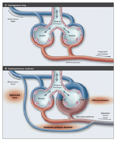
The hepatopulmonary syndrome (HPS) is a triad of liver disease, increased alveolar-arterial oxygen gradient, and intrapulmonary vascular dilatations.

set of four diagnostic criteria for the hepatopulmonary syndrome:

  1. Presence of chronic hepatic disease (alcoholic, post-necrotic, or primary biliary cirrhosis or active chronic hepatitis). Severe liver dysfunction may not be mandatory.
  2. Absence of intrinsic cardiopulmonary disease, with normal chest radiograph or with nodular basal shadowing.
  3. Pulmonary gas exchange abnormalities. An increased alveolar-arterial oxygen gradient (>/= 20 mm Hg) with or without hypoxemia.
  4. The extrapulmonary appearance of IV radiolabeled microspheres or a positive contrast-enhanced echo-cardiogram, suggesting intrapulmonary vascular abnormalities.
  • The unique striking pathological feature of hepatopulmonary syndrome is gross dilatation of the pulmonary precapillary and capillary vessels (to 15 to 100 µm in diameter when the patient is at rest), coupled with an absolute increase in the number of dilated vessels visualized by means of injection at autopsy. hps2

In addition, a few pleural and pulmonary arteriovenous communications (shunts) and portopulmonary venous anastomoses can be seen.

The prerequisite of pulmonary vascular dilatation facilitates the passage of mixed venous blood either rapidly or even directly, through intrapulmonary shunt, into the pulmonary veins.Because of gravity, shifting of blood to the dilated precapillary beds of the lung bases results in an increased hypoxemic dyspnea when the patient is in the upright position.

Ventilation–perfusion mismatch emerges as the predominant mechanism.


Friday, October 3, 2008

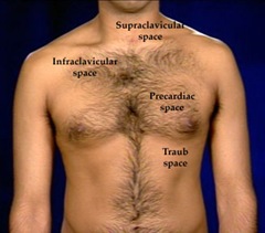
Traube's space

It's a crescent-shaped space overlying the stomach.

The surface markings for Traube’s space traube space

  • the left sixth rib,
  • the left mid-axillary line and
  • the left costal margin.

Percussion should be carried out at one or more levels of Traube’s space from medial to lateral.

Anatomical boundaries are:

1. Right : the inferior margin of the left lobe of  liver.
2. Left : the anterior border of the Spleen.
3. Superior : lower edge of the left lung (Resonance of lung).
4. Inferior : Costal margin.

Contents

1. Fundus of stomach (Hence percussion of Traubes area normally gives Tympanic note).
2. Costo-phrenic recess of left pleura devoid of lungs.

  • Underneath lies the stomach, which produces a tympanic sound on percussion (medicine). If percussion over Traube's space produces a dull tone, this indicates splenomegaly
  • This maneuver has a sensitivity and specificity between 60 and 70% for splenetic enlargement; however, the sensitivity and specificity increases to approximately 80% in non-obese patients who are fasting.

Other Causes of dull sound on percussion of Traube's space:
1. Full stomach.
2. Left sided Pleural effusion.
3. Enlargement of left lobe of liver due to any etiology.
4. Dextrocardia.
5. Proliferative growth in fundus of stomach.

Note: A left lung mass lesion/consolidation alone never produces impairment as lung is not extending to traube's space.

Tuesday, September 23, 2008

Marfan's Syndrome

Clinical signs & diagnostic evaluation of Marfan's syndrome with illustrations.

Download the most clinically useful pdf file from 4shared.

Saturday, August 30, 2008

Syndrome X

Syndrome X or Metabolic syndrome:

Metabolic syndrome is also known as metabolic syndrome X, syndrome X, insulin resistance syndrome, Reaven's syndrome

The metabolic syndrome consists of multiple, interrelated risk factors of metabolic origin that appear to directly promote the development of atherosclerotic cardiovascular disease (ASCVD). This constellation of metabolic risk factors is strongly associated with type 2 diabetes mellitus or the risk for this condition.

Diagnostic criteria

Measure (Any 3 of 5 Criteria Constitute Diagnosis of Metabolic Syndrome) Categorical Cut Points
Elevated waist circumference

≥102 cm (≥40 inches) in men

≥88 cm (≥35 inches) in women

Elevated Blood Pressure

≥130 mm Hg systolic BP

or

≥85 mm Hg diastolic BP

or

Drug treatment for hypertension

Elevated fasting glucose

≥100 mg/dL

or

Drug treatment for elevated glucose

Elevated Triglycerides

≥150 mg/dL (1.7 mmol/L)

or

Drug treatment for elevated TG

Reduced HDL-Cholesterol

<40 mg/dL (1.03 mmol/L) in men

<50 mg/dL (1.3 mmol/L) in women

or

Drug treatment for reduced HDL-C


note:

  • To measure waist circumference, locate top of right iliac crest. Place a measuring tape in a horizontal plane around the abdomen at level of iliac crest. Before reading tape measure, ensure that tape is snug but does not compress the skin and is parallel to floor. Measurement is made at end of normal expiration.
  • Lower waist circumference cut point (eg, ≥90 cm [35 inches] in men and ≥80 cm [31 inches] in women) appears to be appropriate for Asians.
  • Fibrates and nicotinic acid are the most commonly used drugs for elevated Triglycerides and reduced HDL-C. Patients taking 1of these drugs presumed to have high Triglycerides and low HDL.
  • Abdominal obesity is highly correlated with and easier to measure than other indicators of insulin resistance and as  abdominal obesity incorporates both concepts of obesity and insulin resistance as being the 2 major underlying risk factors of the metabolic syndrome

The primary goal of clinical management of the metabolic syndrome is to reduce risk for clinical atherosclerotic disease.

A closely related goal is to decrease the risk for type 2 diabetes mellitus in those patients who do not yet manifest clinical diabetes.


Syndrome Z = Metabolic Syndrome X + obstructive sleep apnea

Other entities which are also known as Syndrome X:

  1. cardiac syndrome X
  2. Fragile X syndrome

Cardiac syndrome X:

  • Cardiac syndrome X occurs when a patient has all of the symptoms of angina pectoris without coronary artery disease or spasm.

note:Prinzmetal angina or variant angina occurs as a result of transient coronary artery spasms. These spasms can occur either at rest or with exertion.

  • Cardiac syndrome X, the triad of
  1. angina pectoris,
  2. a positive exercise electrocardiogram for myocardial ischaemia and
  3. angiographically smooth coronary arteries
  • The main cause of SX is coronary microvascular dysfunction, as indicated by an abnormal response of coronary microcirculation to both vasoconstrictor and vasodilatory stimuli (microvascular angina)

Fragile X syndrome:

  • the most common form of inherited mental retardation
  • the second-leading cause of genetically associated mental retardation after Down syndrome

cause:

The genetic defect is dynamic and lies at the distal end of the long arm of the X chromosome.

Careful examination of the karyotype of affected individuals' lymphocytes, cultured in a folate-depleted and thymidine-depleted medium, reveals a constriction followed by a thin strand of genetic material that extends beyond the long arm at the highly conserved band Xq27.3.

This constriction and thin strand produce the appearance of a fragile portion of the X chromosome, leading to the term fragile X.

The function of fragile X mental retardation (FMR1) gene is believed to play a role in normal brain development.

contains a repeating base pair triplet (CGG) expansion, which is responsible for fragile X syndrome.

features:

  • The phenotype of fragile X syndrome is difficult to diagnose in prepubertal children. Most physical examination findings are notable only after onset of puberty.

150px-Fragile_x_syndrom

    • Growth: Childhood growth is marked by an early growth spurt. However, adult height is often average or slightly below average.
    • Craniofacial: Adolescent and adult patients have a long, thin face with prominent ears, facial asymmetry, a head circumference higher than the 50th percentile, and a prominent forehead and jaw.
  • Ears: Ears are typically large and may protrude.
  • Genitals: Macroorchidism is universal in adult males. In unaffected males, average testicular volume is 17 mL; in patients with fragile X syndrome, testicular volume is more than 25 mL and can be as high as 120 mL.

other physical characteristics can include:

  • Mouth: The mouth has dental overcrowding and a high-arched palate.
  • Eyes: Strabismus is frequently noted.
  • Extremities: Hands and feet manifest nonspecific findings, including hyperextensible finger joints, hand calluses, double-jointed thumbs, a single palmar crease, and pes planus.
  • Back and chest: Pectus excavatum and scoliosis are frequent findings.
  • Cardiac: A heart murmur or click consistent with mitral valve prolapse is often auscultated and requires consultation with a cardiologist.
  • Cognitive history
    • IQ in males frequently indicates mild-to-severe mental retardation (20-70). Females and less-affected males may have IQs that approach 80.