Pages

Showing posts with label clinical skills. Show all posts

Sunday, August 16, 2009

Eponymous Signs in Splenic rupture

Ballance's  sign - Ballance's sign is dullness to percussion in the left flank LUQ and shifting dullness to percussion in the right flank seen with splenic rupture/hematoma. The dullness in the left flank is due to coagulated blood, the shifting dullness on the right due to fluid blood.

Kehr's  sign - Kehr's sign is the occurrence of acute pain in the tip of the shoulder due to the presence of blood or other irritants in the peritoneal cavity when a person is lying down and the legs are elevated. Kehr's sign in the left shoulder is considered a classical symptom of a ruptured spleen. Kehr's sign is a classical example of referred pain: irritation of the diaphragm is signalled by the phrenic nerve as pain in the area above the collarbone.

Saegasser's  sign  - Palpation of  left upper quadrant  inferior to ribs  elicits neck pain in the  patient. 

Wednesday, July 8, 2009

Splitting of Second heart sound

To understand Splitting of S2 better, we should first understand its normal physiology.

Second heart sound:

It has two audible components, the aortic valve closure sound (A2) and the pulmonic valve closure sound (P2), which are normally split on inspiration and virtually single on expiration.

Technique:

It has two audible components, the aortic closure sound (A2) and the pulmonic closure sound (P2), which must be separated by more than 20 msec (0.20 sec) in order to be differentiated and heard as two distinct sounds. It is clinically very important to determine the presence and degree of respiratory splitting and the relative intensities of A2 and P2.

Splitting is best identified in the second or third left ICS, since the softer P2 normally is confined to that area, whereas the louder A2 is heard over the entire precordium, including the apex.

Physiology:

Various research findings suggests that closure of the aortic and pulmonic valves initiates the series of events that produces the second heart sound. The main audible components, however, result from vibrations of the cardiac structures after valve closure. Using high-fidelity, catheter-tipped micro-manometers and echophonocardiography, it has been shown that the aortic and pulmonic valves close silently and that co-aptation of the aortic valve cusps precedes the onset of the second sound by a few milliseconds. The second sound therefore originates from after-vibrations in the cusps and in the walls and blood columns of the great vessels and their respective ventricles. The energy from these oscillations comes from sudden deceleration of retrograde flow of the column of blood in the aorta and pulmonary artery when the elastic limits of the tensed valve leaflets are met. This abrupt deceleration sets the whole cardiohemic system into vibration.

In order to understand splitting of the second heart sound, knowledge of its relationship to the cardiac cycle is essential.

First lets understand why is A2 before P2 ?

1.Both right & left ventricular systole ends at the same time .(ie, systolic muscle contraction of both ventricles ends at same time)

2.pulmonary arterial pressure is less than aortic pressure (i.e, pulmonary resistance to forward flow from ventricles is less than aortic resistance => therefore we can say that pulmonary impedance is less than aortic impedance.){impedance is nothing but resistance}

3.Therefore as pulmonary impedance is less, even after right ventricular systolic contraction blood continues to flow through valve until pulmonary arterial pressure increases more than right ventricle). But as aortic impedance is more ,it stops blood flow through the aortic valve before itself.

4.Due to the above reasons ,Right ventricular ejection begins prior to left ventricular ejection, has a slightly longer duration, and terminates after left ventricular ejection, resulting in P2 normally occurring after A2.

A2 and P2 are coincident with the incisura of the aorta and pulmonary artery pressure curves, respectively, and terminate left and right ventricular ejection periods.(incisura reflects closure of valves)

The differences between the aortic and pulmonary artery vascular impedance characteristics are also essential to understanding the effects of respiration on splitting of S2. When the pressure curves of the pulmonary artery and right ventricle are recorded simultaneously, the pulmonary artery curve at the level of the incisura (dicrotic notch) lags behind the right ventricular curve, or "hangs out" after it.

hangout interval

The duration of the "hangout interval" is a measure of impedance in the pulmonary artery system. In the highly compliant (low-resistance, high-capacitance) pulmonary vascular bed, the hangout interval may vary from 30 to 120 msec, contributing significantly to the duration of right ventricular ejection.

In the left side of the heart, because impedance is much greater, the hangout interval between the aorta and left ventricular pressure curves is negligible (less than or equal to 5 msec).

The hangout interval therefore correlates closely with impedance of the vascular bed into which blood is being injected. Its duration appears to be inversely related to vascular impedance.

Normal physiological splitting during respiration:

Alterations in the impedance characteristics of the pulmonary vascular bed and the right-sided hangout interval are responsible for many of the observed changes in splitting of S2.

In a normal physiologic setting, inspiration lowers impedance in the pulmonary circuit, prolongs the hangout interval and delays pulmonic valve closure, resulting in audible splitting of A2 and P2.

On expiration, the reverse occurs: pulmonic valve closure is earlier, and the A2–P2 interval is separated by less than 30 msec and may sound single to the ear. Since the pulmonary circulation has a much lower impedance than the systemic circulation, flow through the pulmonic valve takes longer than flow through the aortic valve. The inspiratory split widens mainly because of delay in the pulmonic component.

Traditionally it was believed that an inspiratory drop in intrathoracic pressure favored greater venous return to the right ventricle, pooling of blood in the lungs, and decreased return to the left ventricle. The increase in right ventricular volume prolonged right-sided ejection time and delayed P2; the decrease in left ventricular volume reduced left-sided ejection time and caused A2 to occur earlier. The delayed P2 and early A2 associated with inspiration, however, are best understood as an interplay between changes in the pulmonary vascular impedance and changes in systemic and pulmonary venous return. The net effect is that right ventricular ejection is prolonged, left ventricular ejection is shortened, and the A2–P2 interval widens during inspiration.

Clinical Significance

Normally the aortic closure sound (A2) occurs prior to the pulmonic closure sound (P2), and the interval between the two (splitting) widens on inspiration and narrows on expiration. With quiet respiration, A2 will normally precede P2 by 0.02 to 0.08 second (mean, 0.03 to 0.04 sec) with inspiration. In younger subjects inspiratory splitting averages 0.04 to 0.05 second during quiet respiration. With expiration, A2 and P2 may be superimposed and are rarely split as much as 0.04 second. If the second sound is split by greater than 0.04 second on expiration, it is usually abnormal.

Therefore, the presence of audible splitting during expiration (i.e., the ability to hear two distinct sounds during expiration) is of greater significance at the bedside in identifying underlying cardiac pathology than is the absolute inspiratory increase in the A2–P2 interval.

ABNORMAL SPLITTING OF THE SECOND HEART:

(1) persistently single;

(2) persistent (audible expiratory) splitting, with normal respiratory variation;

(3) persistent splitting without respiratory variation (fixed splitting); and

(4) reversed (paradoxical) splitting.

splitting s2

(1) persistently single;

  • When S2 remains single throughout the respiratory cycle, one component is absent or the two components are persistently synchronous.
  • The most common cause of a single S2 is inaudibility of the P2 in older adults with increased anteroposterior chest dimensions.
  • In the setting of
    congenital heart disease, a single S2 due to absence of the pulmonary component is a feature of pulmonary atresia, severe pulmonary valve stenosis, dysplastic pulmonary valve, or complete transposition of the great arteries.
  • Conversely, a single S2 due to inaudibility of the A2 occurs when the aortic valve is immobile (severe calcific aortic stenosis) or atretic (aortic atresia).

(2) persistent (audible expiratory) splitting, with normal respiratory variation;

  • Persistent splitting may be due to a delay in P2, as in cases of simple complete right bundle branch block,or to early timing of the A2, as occasionally occurs in cases of mitral regurgitation(since early emptying of left ventricle –> early closure of Aortic valve).
  • Normal directional changes in the interval of the split (greater with inspiration, lesser with exhalation) in the presence of persistent audibility of both components defines the split as persistent but not fixed.
  • these conditions have wide inspiratory split

(3) fixed splitting;

  • This term applies when the interval between the A2 and P2 is not only wide and persistent but also remains unchanged during the respiratory cycle.
    Fixed splitting is an auscultatory hallmark of  atrial septal defect(ASD). 
  • A2 and P2 are widely separated during exhalation and exhibit little or no change in the degree of splitting during inspiration.

Reason behind wide splitting:

The wide splitting is caused by a delay in the P2 because a marked decrease in pulmonary vascular impedance prolongs the interval between the descending limbs of the pulmonary arterial and right ventricular pressure pulses (“hangout”), and therefore delays the pulmonary incisura and the P2.

Reason behind fixed splitting:

  • We know that in normal individuals , the amount of pulmonary ejection is more during inspiration compared to that in expiration (since pulmoary impedance decreases during inspiration)
  • In ASD .the amount of blood which passes throught he pulmonary valve ramains the samee in both inspiration& expiration because of Phasic changes in systemic venous return during respiration in patients with atrial septal defect are associated with reciprocal changes in the volume of the left-to-right shunt, minimizing respiratory variations in right ventricular filling. (ie, whenever venous return increases in inspiration, it causes a reciprocal decrease in left to right shunting in atria & whenever venous return decreased as in expiration ,shunting increases =>this maintains equal amount of blood in right ventricle irrespective of inspiration or exoiration)
  • The net effect is the characteristic wide, fixed splitting of the two components of the S2.

(4) reversed (paradoxical) splitting. 

  • This term refers to a reversed sequence of semilunar valve closure, the P2 preceding the A2.
  • Common causes of paradoxical splitting are complete left bundle branch block or a right ventricular pacemaker, both of which are associated with initial activation of the right side of the ventricular septum, and delayed activation of the left ventricle owing to transseptal (right-to-left) depolarization.
  • When the S2 splits paradoxically, its two components separate during exhalation and become
    single (synchronous) during inspiration .
  • Inspiratory synchrony is achieved as the two components fuse because of a delay in the P2, less to earlier timing of the aortic
    component.

  splitting s2


Please review this article & if you have any doubts do tell me.

Thursday, July 2, 2009

Brainstem Rules of 4


In 2005, Peter Gates published a superb paper titled:

‘The rule of 4 of the brainstem: a simplified method for understanding brainstem anatomy and brainstem vascular syndromes for the non-neurologist’.

Gates described a simplified method for answering the question ‘Where is the lesion?’ using only the parts of the brainstem that we actually examine during a clinical examination to understand brainstem vascular syndromes.

Firstly, a quick review of the blood supply of the brainstem. Simply put the blood supply comes from:

  1. paramedian branches
  2. long circumferential branches (SAP)
    • superior cerebellar artery (SCA)
    • anterior inferior cerebellar artery (AICA)
    • posterior inferior cerebellar artery (PICA)

And occlusion of these two groups of vessels results in two distinct types of brainstem syndrome:

  1. medial (or paramedian) brainstem syndromes ( due to para-median branch occlusion)
  2. lateral brainstem syndromes ( due to occlusion of the circumferential branches, also occasionally seen in unilateral vertebral occlusion) blood2

And now the rules. If you can remember these rules the  diagnosis of brainstem vascular syndromes becomes a pitifully simple exercise (?!) - here’s how it works:

In the rule of 4 there are 4 rules

  1. There are 4 structures in the ‘midline‘ beginning with M
  2. There are 4 structures to the ‘side‘ (lateral) beginning with S
  3. There are 4 cranial nerves in the medulla, 4 in the pons and 4 above the pons (2 in the midbrain)
  4. The 4 motor nuclei that are in the midline are those that divide equally into 12 except for 1 and 2, that is 3, 4, 6 and 12
    (5, 7, 9 and 11 are in the lateral brainstem)

The 4 medial structures and the associated deficits are:

  1. Motor pathway (or corticospinal tract):
    contralateral weakness of the arm and leg
  2. Medial Lemniscus:
    contralateral loss of vibration and proprioception in the arm and leg
  3. Medial longitudinal fasciculus:
    ipsilateral inter-nuclear ophthalmoplegia
    (failure of adduction of the ipsilateral eye towards the nose and nystagmus in the opposite eye as it looks laterally)
  4. Motor nucleus and nerve:
    ipsilateral loss of the cranial nerve that is affected (3, 4, 6 or 12)

The 4 ’side’ (lateral) structures and the associated deficits are:

  1. Spinocerebellar pathway:
    ipsilateral ataxia of the arm and leg
  2. Spinothalamic pathway:
    contralateral alteration of pain and temperature affecting the arm, leg and rarely the trunk
  3. Sensory nucleus of the 5th cranial nerve:
    ipsilateral alteration of pain and temperature on the face in the distribution of the 5th cranial nerve
    (this nucleus is a long vertical structure that extends in the lateral aspect of the pons down into the medulla)
  4. Sympathetic pathway:
    ipsilateral Homer’s syndrome, that is partial ptosis and a small pupil (miosis)

According to Gates:

These pathways pass through the entire length of the brainstem and can be likened to ‘meridians of longitude‘ whereas the various cranial nerves can be regarded as ‘parallels of latitude‘. If you establish where the meridians of longitude and parallels of latitude intersect then you have established the site of the lesion.

The 4 cranial nerves in the medulla are CN9-12:

  1. Glossopharyngeal (CN9):
    ipsilateral loss of pharyngeal sensation
  2. Vagus (CN10):
    ipsilateral palatal weakness
  3. Spinal accessory (CN11):
    ipsilateral weakness of the trapezius and stemocleidomastoid muscles
  4. Hypoglossal (CN12):
    ipsilateral weakness of the tongue

The 12th cranial nerve is the motor nerve in the midline of the medulla. Although the 9th, 10th and 11th cranial nerves have motor components, they do not divide evenly into 12 (using our rule) and are thus not the medial motor nerves.

The 4 cranial nerves in the pons are CN5-8:

  1. Trigeminal (CN5):
    ipsilateral alteration of pain, temperature and light touch on the face back as far as the anterior two-thirds of the scalp and sparing the angle of the jaw.
  2. Abducent (CN6):
    ipsilateral weakness of abduction (lateral movement) of the eye (lateral rectus).
  3. Facial (CN7):
    ipsilateral facial weakness.
  4. Auditory (CN8):
    ipsilateral deafness.

The 6th cranial nerve is the motor nerve in the medial pons.
The 7th is a motor nerve but it also carries pathways of taste, and using the rule of 4 it does not divide equally in to 12 and thus it is not a motor nerve that is in the midline.
The vestibular portion of the 8th nerve is not included in order to keep the concept simple and to avoid confusion. Nausea and vomiting and vertigo are often more common with involvement of the vestibular connections in the lateral medulla.

The 4 cranial nerves above the pons are CN1-4:

  1. Olfactory (CN1):
    not in midbrain.
  2. Optic (CN2):
    not in midbrain.
  3. Oculomotor (CN3):
    impaired adduction, supradduction and infradduction of the ipsilateral eye with or without a dilated pupil.
    The eye is turned out and slightly down.
  4. Trochlear (CN4):
    eye unable to look down when the eye is looking in towards the nose (superior oblique).

The 3rd and 4th cranial nerves are the motor nerves in the midbrain.

Thus a medial brainstem syndrome will consist of the 4 M’s and the relevant motor cranial nerves, and a lateral brainstem syndrome will consist of the 4 S’s and either
the 9-11th cranial nerve if the lesion is in the medulla, or the 5th, 7th and 8th cranial nerve if the lesion is in the pons.

Handy tip:
If there are signs of both a lateral and a medial (paramedian) brainstem syndrome, then one needs to consider a basilar artery problem, possibly an occlusion.

I’ll let you mull over these rules until the next ‘brainstem’ post, where you’ll be able to test drive ‘Gates’ Brainstem Rules of 4′ on some clinical scenarios.

Scenario 1

  • You are examining a patient with sudden onset left-sided weakness. These are your clinical examination findings:
    weakness of the left upper and lower limbs, with sparing of the face.
    tongue deviation to the right, with no ophthalmoplegia.
    loss of vibration and proprioception in the left upper and lower limbs.
  • medial_brainstem_synd
  • Where is the lesion?
    weakness of the left upper and lower limbs, with sparing of the face:
    motor (corticospinal pathway) localises the lesion to the contralateral medial brainstem
    (sparing of the face (CN7) means the lesion must be below the upper pons)
    tongue deviation to the right, with no ophthalmoplegia:
    tongue deviation indicates CN12 involvement, localising the lesion to the ipsilateral medulla
    (sparing of CN3 and CN6 means the midbrain and pons are not involved)
    loss of vibration and proprioception in the left upper and lower limbs:
    confirms localisation of the lesion to the contralateral medial brainstem
  • Site of the lesion: right medial medulla. Sometimes, due to the peculiar pattern of blood supply to the medulla, bilateral infarction may occur.

Scenario 2

  • You are examining a patient with sudden onset right-sided weakness. These are your clinical examination findings:
    weakness of the right face, upper and lower limbs.
    the left eye is turned “down and out” and the pupil is dilated.
  • Where is the lesion?
    weakness of the right face, upper and lower limbs:
    motor (corticospinal pathway) localises the lesion to the contralateral medial brainstem
    (involvement of the face means the lesion must be at or above the upper pons)
    the left eye is turned “down and out” and the pupil is dilated:
    CN3 involvement, localising the lesion to the ipsilateral midbrain
    (sparing of CN6 and CN12 means the pons and medulla are not involved)
  • Site of the lesion: left medial midbrain. A CN3 palsy (from damage to the CN3 nerve fascicle) and contralateral hemiplegia is known as Weber’s syndrome (”basal” infarction)  - which can be difficult to distinguish from ‘coning’ if you don’t have a CT scanner available.

Scenario 3

  • You are examining a patient with vertigo, vomiting, and nystagmus. These are your clinical examination findings:
    left-sided limb ataxia.
    left-sided alteration of pain and temperature on the face.
    left-sided ipsilateral Homer’s syndrome.
    right-sided alteration of pain and temperature affecting the arm and leg.
    dysarthria and decreased gag reflex on the left, with the palate pulling up on the right-side.
  • wallenberg syndrome
  • Where is the lesion?
    left-sided limb ataxia:
    spinocerebellar pathway localises the lesion to the ipsilateral lateral brainstem.
    left-sided alteration of pain and temperature on the face:
    Sensory nucleus of the 5th cranial nerve localises the lesion to the ipsilateral lateral brainstem.
    left-sided ipsilateral Homer’s syndrome:
    Sympathetic pathway localises the lesion to the ipsilateral lateral brainstem.
    right-sided alteration of pain and temperature affecting the arm and leg:
    Spinothalamic pathway localises the lesion to the contralateral lateral brainstem.
    dysarthria and decreased gag reflex on the left, with the palate pulling up on the right-side:
    localises the lesion to the medulla affecting the ipsilateral  CN9 and 10.
  • Site of the lesion: left lateral medulla. Also known as Wallenberg’s syndrome, caused by a left vertebral or left posterior inferior cerebellar artery occlusion (blood supply is variable to this region).

Scenario 4

  • You are examining a patient with right-sided deafness, that was preceded by tinnitus. These are your clinical examination findings:
    right-sided limb ataxia (predominantly affecting the right upper limb).
    right-sided facial numbness with loss of the corneal reflex.
    right-sided hemi-facial spasms.
  • Where is the lesion?
    right-sided limb ataxia (predominantly affecting the right upper limb):
    spinocerebellar pathway localises the lesion to the ipsilateral lateral brainstem.
    right-sided facial numbness with loss of the corneal reflex:
    Sensory nucleus of the 5th cranial nerve localises the lesion to the ipsilateral lateral brainstem.
    right-sided hemi-facial spasms:
    the lesion involves the pons affecting the ipsilateral CN7.
  • Site of the lesion: The findings indicate a lesion affecting the right lateral pons with evidence of spinocerebellar involvement. In this case the lesion was not vascular in origin but in fact an example of a cerebropontine angle lesion - an acoustic neuroma (or schwannoma). This demonstrates the the broader utility of Gates’ Brainstem Rules of 4.

Scenario 5

  • You are examining a patient with sudden onset right-sided weakness. These are your clinical examination findings:
    weakness of the right face, upper and lower limbs.
    failure of abduction of the left eye.
    loss of vibration and proprioception in the right upper and lower limbs.
  • Where is the lesion?
    weakness of the right face, upper and lower limbs:
    motor (corticospinal pathway) localises the lesion to the contralateral medial brainstem
    (involvement of the face means the lesion must be at or above the upper pons)
    failure of abduction of the left eye:
    indicates CN6 involvement, localising the lesion to the ipsilateral pons.
    (sparing of CN3 and CN12 means the midbrain and medulla are not involved)
    loss of vibration and proprioception in the right upper and lower limbs:
    confirms localisation of the lesion to the contralateral medial brainstem
  • Site of the lesion: left medial pons. Interestingly, the facial nerve runs a strange course - it loops around medial to the CN6 nucleus from its own laterally situated CN7 nucleus. Thus a CN7 palsy tends to coexist with a CN6 lesion despite the CN7 nucleus being in the lateral pons.

Scenario 6

  • You are examining a patient with sudden onset intermittent double vision (diplopia). These are your clinical examination findings:
    failure of adduction past the midline (movement towards the nose) of the left eye and leading eye (right) nystagmus on looking laterally to the right. Normal eye movements on looking to the left.
    The patient is hypertensive. There is no hemiparesis and further examination is unremarkable.
  • Where is the lesion?
    This finding  suggests a unilateral left-sided internuclear ophthalmoplegia, which localises the lesion to the ipsilateral medial longitudinal fasciculus (MLF). The MLF connects CN3 in the midbrain and the contralateral CN6 in the pons.
    The MLF is not usually affected when there is hemiparesis as it lies further back in the brainstem relative to the motor (corticospinal) pathway.
    Unilateral internuclear ophthalmoplegia can result from a lacunar infarct.
    (Always remember the possibility of multiple sclerosis)
  • Site of the lesion: left medial longitudinal fasciculus (connects CN3 in the midbrain and contralateral CN6 in the pons).

Scenario 7

  • You are examining a patient with a right-sided Horner’s syndrome. These are your clinical examination findings:
    right-sided Horner’s syndrome.
    right-sided limb ataxia.
    left-sided total loss of sensation.
  • Where is the lesion?
    right-sided Horner’s syndrome:
    sympathetic pathway localises the lesion to the ipsilateral lateral brainstem.
    right-sided limb ataxia:
    spinocerebellar pathway localises the lesion to the ipsilateral lateral brainstem.
    left-sided total loss of sensation:
    Spinothalamic pathway localises the lesion to the contralateral lateral brainstem (in the midbrain the medial lemniscal pathway is actually situated more laterally, ventral to the spinothalamic pathway - ie. the two pathways come together… an exception to the Rule of 4!).
  • Site of the lesion: Right dorsolateral midbrain. An extensive lesion that also involves CN3 is known by the delightful name of Nothnagel’s syndrome.

Scenario 8

  • You are examining a patient with a ‘down and out’ right eye with pupillary dilatation. These are your clinical examination findings:
    right-sided impaired adduction, supradduction and infradduction of the ipsilateral eye with a dilated pupil.
    left-sided limb ataxia.
  • Where is the lesion?
    right-sided impaired adduction, supradduction and infradduction of the ipsilateral eye with a dilated pupil:
    CN3 lesion localises the lesion to the ipsilateral medial midbrain.
    left-sided limb ataxia:
    usually this indicates ipsilateral spinocerebellar pathway involvment (a lateral structure). However, in this case we know that the midbrain is affected (CN3 palsy) and the red nucleus lies in the medial midbrain just lateral to the CN3 nerve fascicle. Damage to the red nucleus interrupts the ‘dentatorubrothalamic tract’ from the opposite cerebellar hemisphere causing cerebellar signs in the limbs opposite to the CN3 lesion.
  • Site of the lesion: Right medial midbrain. The clinical manifestations of this lesion (affecting the CN3 nucleus or its fascicle as well as the red nucleus) is known as Benedikt’s syndrome.


Taken from: Life in the Fast Lane

Wednesday, October 1, 2008

Breath sounds made easy.

respiratory-system

To understand breath sounds ,we should first stress on the Anatomy & Physiology of lungs.This is important as we have to know where & how & why these sounds are produced.Don't forget to check the Audio links at the end of the post.

Lobes of Lung & their surface markings:

The right & left lung are divided by deep fissures into lobes.

Right lung Left lung
  • 3 lobes
  • 2 lobes
  • 2 fissures: oblique& horizontal
  • only oblique fissure
  • oblique fissure separates the lower lobe from upper& middle lobe.
  • horizontal fissure between upper& middle lobe.
  • oblique fissure separates the upper& lower lobe.

 lung 3d


Surface markings:

This is very important for us to localize the lesion in lung.

Anterior lung markingsposterior lung markingsright lung markingsleft lung marking

 Areas of Auscultation:

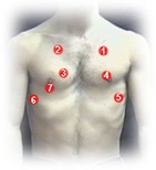
areas of auscultation in frontareas of auscultation back


Breath Sounds:

Normal

Abnormal Adventitious
Tracheal Absent/decreased normal breath sounds Crackles (rales)
Bronchial

Bronchial sounds in abnormal areas.

Wheeze
Bronchovesicular Rhonchi
Vesicular

Normal Breath Sounds

            To be able to distinguish between types of abnormal breath sounds and know their location, it is important to understand normal respiration and its effect on airway noises that make up breath sounds.

  • Normal respiratory cycle

respiraory cycle

  • The inspiratory phase is shorter with faster airflow.
  • The majority of breath sounds, both normal and adventitious, are in the range of 16 to 200 Hz. This is at the lower end of the spectrum of normal hearing (16 to 16,000 Hz).
  • The sensitivity of the human ear in detecting low-frequency sound is relatively poor; thus, breath sounds are difficult to appreciate even with the stethoscope.


Normal Breath Sounds Normal Location of Sound Sound Quality Distinguishing Characteristic Diagram
Tracheal Only heard over the trachea. loud, harsh like air is being blown through a pipe. Expiration slightly longer than inspiration with similar loudness & a pause between. tracheal sound
Bronchial present over the large airways & thus heard over the body of the sternum these sounds are more tubular and  high pitched than vesicular sounds, but not as harsh as tracheal breath sounds.

Expiratory sounds are louder & last longer than inspiratory sounds & have a short pause between inspiration and expiration sounds

bronchial sound
Bronchovesicular heard in the posterior chest between the scapulae and in the first & second intercostal spaces anteriorly. medium pitched Inspiration & expiration sounds equal in length& loudness.these sounds are different from vesicular since they have a pause between inspiration & expiration. bronchovesicular sound
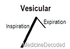
Vesicular heard throughout most of the lung fields( Periphery of lungs) soft, low pitched, or rustling sounds normally Inspiration sound is longer & louder than expiration sound without a pause between them. vesicular sound


You can have a peek into the various sounds for better understanding by pressing the image below.

 breath sounds


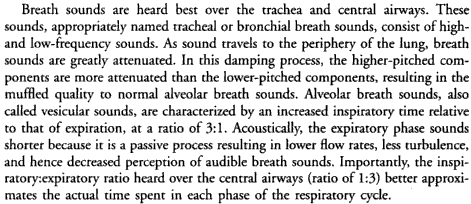
Important :Now you may get the doubt that "why the Vesicular sounds have increased inspiratory time& loudness than expiration sounds which is contradictory to what we have learnt in the beginning of the post about Respiratory cycle?"

The reason is in the below image-

Vesicular sound mechanism

Abnormal Breath sounds:

Abnormal breath sounds

The above so called Normal Breath sounds are considered normal only when they are heard in their respective places ,if they are heard in a different place ,they are abnormal.

Bronchial sounds:

  • When auscultated in other areas, bronchial breath sounds are an abnormal finding.
  • They are heard over areas of consolidation, as solid lung conducts the sound of turbulence in main airways to peripheral areas without filtering.
  • Occasionally breath sounds over a large cavity have an exaggerated bronchial quality. This very hollow or amphoric sound has been likened to that heard when air passes over the top of a hollow jar (Greek amphoreus).  
Causes of bronchial breath sounds( ie, in areas other than over body of sternum)

Common

Lung consolidation (lobar pneumonia)

Uncommon

Localised pulmonary fibrosis

Pleural effusion (above the fluid)

Collapsed lung (eg: adjacent to a pleural effusion)


ADVENTITIOUS SOUNDS:

  • Added lung sounds are divided into two general groups: discontinuous sounds, which are 250 milliseconds or less in duration, and continuous sounds, which last longer than 250 milliseconds .
  • Discontinuous sounds are further classified as either coarse or fine crackles; continuous sounds, as either wheezes & rhonchi.

Crackles

  • explosive, sharp, discrete bursts of interrupted sound.
  • Their pattern is remarkably constant and cannot be destroyed by coughing.
  • Crackles are divided into two types depending on their acoustic properties.
Fine Crackles Coarse Crackles
sounds like the crackling noise made when salt is heated on a frying pan. sound of water being poured from a bottle.
On auscultation fine crackles are in general higher pitched, less intense and of shorter duration than coarse crackles. coarse crackles are in general lower pitched, less intense and of longer duration than fine crackles.
The probable mechanism for the production of fine crackles is as follows. During inspiration, the air pressure on the "upstream" (mouth) side increases until it is able to overcome the forces that are closing the bronchiole. When this occurs, the airway snaps open as the pressure between the bronchiole and the alveolus is equalized . The resulting vibration in the airway causes a discrete, sharp sound of very short duration. The bubbling sound of coarse crackles is produced when air passes over secretions in the larger airways (trachea and bronchi).
Fine crackles are usually appreciated only during inspiration. Since air flows through the airways during inspiration and expiration, coarse crackles are more likely to be detected during both phases of the respiratory cycle.
  The most common conditions associated with coarse crackles are congestive heart failure and pneumonia,Bronchiectasis.
   

  • Consistent with its mechanism ,crackles are typically predominant in lung bases ,where the transpulmonary pressure is lowest & parenchyma less distended.
  • As hundreds of airways open sequentially, the characteristic crackling sound is produced. The sound of fine crackles can be simulated by rubbing a lock of hair between your fingers.

Some clinicians maintain that the timing of onset of crackles aids in the differential diagnosis of parenchymal and airways-disease.

  • Crackles auscultated during early inspiration are thought to be more indicative of airways disease, such as chronic bronchitis, emphysema, and asthma.
  • Crackles auscultated during late inspiration are more suggestive of parenchymal disorders, such as pulmonary fibrosis, interstitial pneumonitis, and pneumonia.
Early inspiratory crackles Mid to Late inspiratory crackles
Dependent atelectasis Bronchiectasis
Bronchitis

Restrictive lung diseases

  • asbestosis
  • idiopathic pulmonary fibrosis
  • sarcoidosis
  • scleroderma lung disease
Asthma pulmonary edema
Emphysema


Continuous:

  1. wheeze
  2. rhonchi

Wheezes

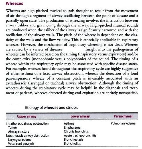
Wheezes are divided according to timing in respiratory cycle & actual sound produced ( monophonic or polyphonic)

Polyphonic wheeze:

  • the most common type
  • typical of COPD & asthma
  • multiple simultaneous different pitched sounds occur during expiration & signify diffuse  airway disease

Fixed monophonic wheeze:

  • note of single pitch resulting from narrowing of a single airway
  • sound does not change with coughing
  • seen in tumor or foreign body case

Sequential inspiratory wheeze:SQUAWKS

  • caused by vibration after opening of previously closed airway
  • typical of extrinsic allergic alveolitis

Stridor:

  • Stridor is a continuous, high-pitched monophonic sound heard throughout respiration; this sound is accentuated during inspiration.
  • implies local obstruction to extra-thoracic airways (which tend to collapse on inspiration)
  • often implies carcinoma or foreign body in major airways

Rhonchi:

books


Pleural rub:

  • The visceral and parietal pleurae normally move silently against each other during respiration. However, when the pleurae are inflamed, the two thickened surfaces produce vibrations as they move irregularly over each other.
  • A pleural rub is the sound produced by the motion of inflamed pleurae. It tends to be a loud, grating sound confined to a relatively small area of the chest wall. A pleural rub is usually heard during inspiration and expiration.
  • A pleural rub has also been described as a leathery sound. Usually, the inspiratory and expiratory components of the rub can be readily heard.
  • When effusion separates the two pleural surfaces, the rub may disappear. However, the disappearance of a pleural rub does not necessarily mean that the pleural inflammation itself has resolved.

A pericardial rub usually has three components (atrial systole, ventricular systole, and ventricular diastole); it can usually be distinguished from a pleural rub by having the patient hold his breath. During breath-holding, a pleural rub disappears but a pericardial rub persists.

Mediastinal crunch (Hamman's sign)

  • grating, crunching sound heard in the center of the anterior chest. The sound coincides with the heartbeat and signifies the presence of free air in the mediastinum. The pathogenesis of mediastinal crunch is not clear, however, it may involve compression of the air by the beating heart and the mediastinal structures.
  • heard over the precordium in spontaneous mediastinal emphysema.
  • This sound is heard best over the left lateral position. It has been described as a series of precordial crackles that correlate with the heart beat and not the respirations. Hamman's crunch is caused by pneumomediastinum or pneumopericardium, and is associated with tracheobronchial injury due to trauma, medical procedures (e.g., bronchoscopy) or proximal pulmonary bleb rupture. It is commonly seen in Boerhaave syndrome.

Transmitted voice sounds:

Egophony, bronchophony, and pectoriloquy all refer to auscultatory signs that can be heard over areas of pulmonary consolidation. The pathogenesis of these signs relates to the increased sound transmission through the consolidated lung. This results in transmission of sound-from the larger bronchi through the consolidated lung to the periphery without significant loss in sound quality.

Transmitted voice sounds Location of sound Method of elicitation Reason( since sound travels faster & better in solids, liquids compared to in air)
Egophony over consolidated lung tissue elicited by having the patient say the letter "E' while you listen with the stethoscope. When egophony is present the "E" sounds like "A." Mass/exudate in the lungs enables greater transmission of sound of patient repeating letter "E"
Bronchophony over consolidated lung tissue demonstrated by having the patient say a phrase as you auscultate; "ninety-nine" is the conventional phrase. The sound will be indistinct and muffled over the normal lung. However, the "ninety-nine" will be heard distinctly over the consolidated lung, without loss of clarity. Mass/exudate in the lungs enables greater transmission of sound of patient repeating number "99"
Whispering Pectoriloquy over consolidated lung tissue similar to bronchophony, except it is usually elicited by having the patient say the phrase "one, two, three." The phrase will be muffled and indistinct when you auscultate over the normal lung and clearly audible when you auscultate over an area of consolidation. Mass/exudate in the lungs enables greater transmission of sound of patient whispering


CLINICAL CORRELATIONS

  • The common obstructive diseases of the lung - chronic bronchitis, emphysema, and asthma - can frequently be distinguished solely on the basis of the physical examination.
  • Patients with chronic bronchitis commonly have noisy chests because of crackles and wheezes. The breath sounds are vesicular in nature, however, there is a prolonged expiratory phase, which generally correlates with the degree of obstruction. Breath sounds at the mouth will be heard at normal intensity.
  • Patients with emphysema, on the other hand, present with a relatively quiet chest. Breath sounds are vesicular but significantly reduced in intensity Adventitious sounds are unusual unless bronchitis or asthma is present.
  • The expiratory phase of vesicular breath sounds is similarly prolonged, however, breath sounds heard at the mouth are usually reduced, in contrast to what is normally heard with chronic bronchitis or asthma.
  • Polyphonic Wheezing is the hallmark of clinical asthma & COPD. In patients with mild or early asthma, wheezing is usually heard over the central airways only during expiration. As bronchospasm worsens, the wheezes are heard over the entire chest and in both phases of respiration. The breath sounds are vesicular, and there is a prolonged expiratory phase that tends to correlate with the degree of bronchial obstruction. Breath sounds heard at the mouth are of normal intensity. Interstitial pneumonitis and fibrosis are characterized by the presence of fine inspiratory crackles. With early or mild disease, the crackles are heard at end-inspiration; however, as the disease progresses, they may occupy more of the inspiratory cycle - it is often difficult to hear the underlying breath sounds because of the intensity of the crackles in interstitial fibrosis, but the sounds are vesicular with no prolongation of expiration.
  • Pulmonary consolidation, as in lobar pneumonia, yields bronchial breath sounds over the affected area. Also, coarse or fine crackles may be heard. Expiration is not prolonged unless obstructive disease exists.
  • Left-sided heart failure. Pulmonary congestion associated with left sided heart failure is characterized by transudative fluid in the interstitium and alveoli. The bronchial mucosa may also be swollen.
  • Breath sounds in left-sided heart failure are vesicular, although they sometimes have a prolonged expiratory phase secondary to bronchial mucosal edema. Fine crackles are heard in mild to moderate pulmonary edema, while coarse crackles and polyphonic wheezes occur in severe pulmonary edema.
  • Pleural effusion. Pleural fluid or pleural thickening muffles the transmission of all lung sounds to the periphery. Vesicular sounds are decreased or absent Because the adjacent lung is compressed, bronchial breath sounds are sometimes heard at the area just above the pleural effusion. The reason for this is,the compressed edematous lung, which is immediately above the effusion, acts as a consolidation and causes increased sound transmission.
  • Pneumothorax. With a small or mild pneumothorax, decreased vesicular breath sounds may be heard on the side that is affected. Breath sounds are absent if a more extensive pneumothorax is present.

links to breath sounds audio:

  • Auscultation assistant:The Auscultation Assistant provides heart sounds, heart murmurs, and breath sounds in order to help medical students and others improve their physical diagnosis skills.

auscultation assistant

 basic lung sounds

web3

 rale repository