Pages

Showing posts with label ophthalmology. Show all posts

Saturday, April 11, 2009

Various Lines in Ophthalmology

Arlt's Line :

  • Horizontal line of conjunctival scarring in sulcus subtarsalis of superior palpebral conjunctiva.
  • Seen in Trachoma.

arlt's line

Ehrlich-Turk Line :

Vertical linear deposition of KPs in uveitis.

Ferry's Line :

Corneal epithelial iron line at the edge of filtering blebs of glaucoma surgery.ferry line

Hudson-Stahli Line :

Iron deposition line in the corneal epithelium, which commonly seen in the junction between middle and lower third cornea.
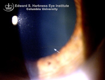
hudson stahli line

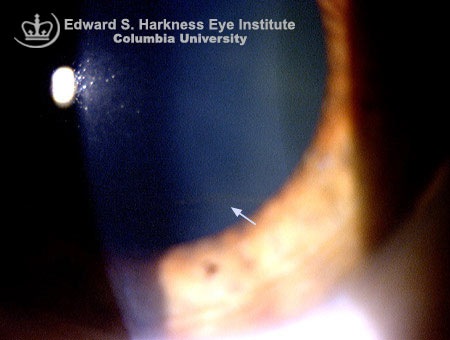
Khodadoust Line :

  • Corneal graft endothelial rejection line composed of inflammatory cells.
  • Khodadoust's line separates immunologically damaged endothelium - usually originating from neovascularization in the same area - from the unaffected endothelium. In the area of damage the endothelium is decompensated resulting in stromal and epithelial edema. Timely immuno-suppression can reverse this process.

Khodadoust's line

Endothelial rejections :Most severe form of corneal rejection as this layer doesn’t have regenerative property & significant cell loss can cause chronic graft edema.

  • Classic endothelial rejection presents with an endothelial rejection line (Khodadoust line) that usually begins at a vascularized portion of the peripheral graft-host junction and progresses, if untreated, across the endothelial surface over several days. The rejection line consists of mononuclear white cells that damage endothelial cells as the line sweeps across the endothelium.
  • Generally, a mild-to-moderate anterior chamber reaction is present. The damaged endothelium is unable to properly dehydrate the corneal graft; as a result, the donor cornea is clear ahead of the rejection line and is cloudy and edematous behind it.
  • A second variant of endothelial rejection is more diffuse in character, with scattered keratic precipitates and an anterior chamber reaction indicative of endothelial rejection and damage. In this type of endothelial rejection, stromal edema typically is not localized, but rather generalized throughout the graft, consistent with the generalized endothelial damage. The combination of keratic precipitates, an anterior chamber reaction, circumcorneal injection, and regions of corneal edema should be diagnosed as corneal graft rejection. In some cases, it may be difficult to distinguish graft edema from rejection and graft edema from endothelial insufficiency. Since rejection may be reversible, it is best in these situations to treat patients as if they have graft rejection.

Paton's Line :

  • Circumferential retinal folds in peripapillary region due to papilledema.
  • Papilledema is defined as optic nerve head edema secondary to increased intracranial pressure. The main cause of optic nerve head swelling is blockage of the axoplasma transport and the blockage occurs at the lamina cribrosa.
  • The optic nerve head can swell to the extent where it is extended forward into the vitreous as well as laterally. This lateral swelling causes the retina to buckle inward at the temporal aspect of the optic nerve head. The buckling is know as Paton's lines or folds.

Paton's Line

Sampaolesi line :

  • Wavy line of Increased pigmentation anterior to Schwalbe's line.
  • Occurs in
    1. pseudoexfoliation syndrome
    2. pigment dispersion syndrome
    3. trauma
    4. iris melanoma

Sampaolesi line

Scheie's Line or Zentmayer's line :

  • Pigment on lens equator and posterior capsule in pigment dispersion syndrome.
  • Peripheral pigmentation of the posterior lens capsule anterior to the junction between the anterior hyaloid face and the posterior lens capsule (ligamentum hyaloideo-capsulare of Wieger)
  • "Scheie's line" is considered to be pathognomonic for pigment dispersion syndrome.

Scheie's Line

Schwalbe's Line :

  • Termination of Descemet's membrane .
  • Line where corneal scleral meshwork terminates anteriorly .Delineates the outer limit of the corneal endothelium layer.

Stockers Line :

Corneal epithelial iron line at the edge of pterygium.

stockers line

White lines of Vogt :

  • Sheathed or sclerosed vessels seen in Lattice degeneration.
  • Lattice degeneration of the retina is a fairly common degenerative disease of the peripheral retina characterized by the presence of lattice lines created by fibrosed blood vessels.
  • Crisscrossing fine white lines that account for the name lattice degeneration are present in roughly only 10% of lesions and most likely represent hyalinized blood vessels.

white lines vogt

Friday, April 10, 2009

Corneal Pigmentation – Decoded


Corneal Pigmentation

Type of Pigment Name of Deposit Disorder Location in Cornea
Iron Stocker’s line Pterygium Epithelium
Hudson-Stahli line In old age Epithelium
Ferry’s line Filtering bleb Epithelium
Fleischer’s ring Keratoconus Epithelium
Coat’s white ring Previous Metallic foreign body injury Bowman’s layer 
Siderosis Stroma
Blood staining of cornea Stroma
Copper Kayser-Fleischer ring Wilsons’s Disease Descement’s membrane
Melanin Krukenberg’s spindle Pigment dispersion syndrome Endothelium
Silver Argyrosis Stroma
Gold Chrysiasis Stroma

Stockers line:

  • Iron deposition line in the corneal epithelium, located at the corneal leading edge of a pterygium.
  • present at the leading edge of non-growing pterygium
  • yellow to golden brown.

stocker's line

Hudson-stahli line:

  • Iron deposition line in the corneal epithelium, which commonly seen in the junction between middle and lower third cornea (corresponding to the line of lid closure).
  • Often seen in elderly.
  • bilateral & symmetric
  • Causes no symptoms or clinical significance.

hudson stahli line

Ferry’s line:

  • Seen in front of filtering bleb in glaucoma.
  • Filtering bleb is a blister of conjunctiva resulting from glaucoma surgery by which a flap of sclera is created in the eye wall, allowing aqueous humor to percolate out of the eye and underneath the conjunctiva, thus lowering intraocular pressure. To know more about it, check this MedRounds link.

ferry's line

Coat's White Ring:

  • A form of iron deposit at the level of Bowman's layer.
  • Clinical features:
    • Asymptomatic
    • Small, granular, oval white ring
    • Commonly associated with previous corneal foreign body injury
  • Requires no treatment.

coats white ring

Fleischer Ring :

  • Partial or complete iron deposition ring in deep epithelium encircling the base of the cone.
  • Formed when hemosiderin (iron) pigment is deposited deep in the epithelium
  • Characteristic in eyes with keratoconus.

fleischer ring

Kayser-Fleischer ring:

  • Deposits of copper producing golden-brown discoloration in the peripheral cornea. Their earliest appearance consists of colored crescents at the superior and inferior quadrants of the cornea that eventually become circumferential.
  • The Kayser-Fleischer ring is the single most important diagnostic sign in Wilson's disease
  • It is found in 95% of patients.
  • Virtually all patients with Kayser-Fleischer rings have neurological manifestations.
  • Diseases associated with Kayser-Fleischer rings
    1. Wilson's disease
    2. primary biliary cirrhosis
    3. chronic cholestasis
    4. cryptogenic cirrhosis

kayser fleischer ring

Krukenberg’s spindle:

  • a vertically oriented deposition of pigment on the posterior endothelial surface of cornea.
  • Seen in Pigment dispersion syndrome.
  • In Pigment dispersion syndrome, the iris is bowed posteriorly causing it to rub against the lens zonules. this results in the loss of pigment epithelium& hence the transillumination and the endothelium deposition of pigment. The vertical orientation of pigment is due to conventional current.
  • Pigment granule accumulation in the trabecular meshwork then leads to progressive trabecular dysfunction and ocular hypertension with or without associated glaucomatous optic neuropathy.About 1/3 of them develop pigmentary glaucoma.
  • The classic triad of clinical signs of PDS consists of a
    1. Krukenberg spindle,
    2. slit-like, radial, mid-peripheral iris transillumination defects, and
    3. pigment deposition on the trabecular meshwork (Sampaolesi’s line)

krukenberg spindlepigment dispersion syndrome

Friday, March 6, 2009

Wonderful set of Neuro-Ophthalmology video lectures- Must watch.

This first lecture covers basic eye muscle anatomy, cranial nerve palsies, and gives a basic approach to localizing cranial nerve lesions.

Neurology in ophthalmology can be quite tricky, so hopefully this video explains a good approach to extraocular eye movements.

This lecture was created and recorded by Dr. Timothy Root, an ophthalmologist with a background in multimedia education.


For the second Video Lecture - click on Read More.

The second lecture covers a few more neurology topics: including visual fields, myasthenia gravis, thyroid eye disease, optic neuritis, neuropathy, and giant cell arteritis.


Thursday, March 5, 2009

Eponyms about Vogt - Ophthalmology

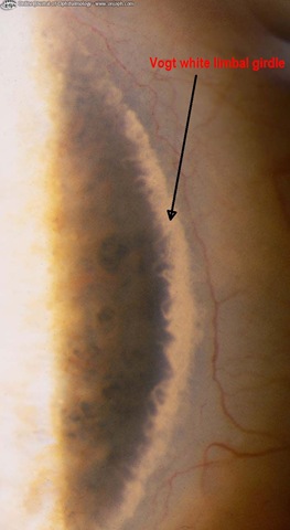
Vogt white limbal girdle:

  • Age related corneal degeneration.
  • Characterized by bilateral, narrow cresentic lines composed of chalky white deposits(present in Bowman’s membrane) in the interpalpebral fissure along the nasal& temporal limbus.

 vogt's white limbal girdle

  • 2 TYPES ARE PRESENT
Type1 Type2
separated from limbus by clear interval No clear zone in between

To continue reading, click on READ MORE.

Vogt's striae :

  • Seen in keratoconus.
  • With thinning of the cornea vertical lines in the Descemet's membrane become visible. They disappear with pressure on the globe.

vogt striae

Vogt's anterior mosaic crocodile shagreen :

  • Age related corneal degeneration.
  • Anterior mosaic crocodile shagreen appears as bilateral, polygonal, grayish-white opacities separated by relatively clear spaces in the deep layers of the epithelium and in Bowman's layer.

vogt crocodile shagreen patches

Vogt-Koyanagi-Harada (VKH) syndrome: Vogt-Koyanagi-Harada (VKH) disease is a multisystem disorder characterized by granulomatous panuveitis with exudative retinal detachments that is often associated with neurologic and cutaneous manifestations.

Cutaneous features Neurological features Ocular features
Alopecia Meningeal involvement Chronic granulomatous iridocyclitis
Poliosis Encephalopathy

Posterior sgment involvement:

  • multifocal choroiditis
  • multifocal detachments of sensory retina
  • exudative retinal detachment
Vitiligo

Auditory symptoms:

  • tinnitus
  • vertigo
  • deafness

Lucid interval of vogt:

  • Seen in Arcus senilis(Age related corneal degeneration)
  • Arcus senilis appears as a single greyish ring compose of lipid deposits parallel to the limbus and separated from it by a 1 mm lucid interval of Vogt.
  • The classic theory concerning the appearance of arcus cornealis is that lipids migrate from the limbal vascular arcade to the peripheral cornea and the clear interval of Vogt is formed due to absorption of the lipids that are closer to the vascular arcade.

lucid interval of vogt

Wednesday, March 4, 2009

Eponyms about Elschnig in Ophthalmology

  • Elschnig's pearls(Elschnig's bodies): Pearl-like clusters formed by growth of Equatorial epithelial cells onto the posterior lens capsule of the eye following cataract surgery. (A type of After cataract)

elschnig pearls Read the remaining collection of eponyms by clicking Read More.

  • Elschnig's conjunctivitis:Chronic conjunctivitis associated with hyperplasia of the tarsal gland and the frothy secretions.
  • Elschnig's spots:In malignant hypertension, blood vessels in Retina& Choroid may be damaged leading to fibrinoid necrosis(presence of fibrin thrombi in vascular lumen). So when choroid vessels are damaged, it produces choroidal infarcts& necrosis of overlying Retinal Pigment Epithelium (since the outer 4 layers of retina are supplied by Choriodal blood vessels).These spots appear as small isolated circular areas having central retinal pigment, epithelial pigment clumping, and a surrounding halo of de-pigmentation.These are known as Elschnig spots.(So seen when choroidal blood vessels damaged in Malignant hypertension-Hypertensive choriodopathy)

elschnig  spots

  • Z-plasty (Elschnig's operation): It is useful to correct mild to moderate degree of cicatricial
    Ectropion.

z plasty(elschnig's operation)

  • Elschnig's syndrome(Elschnig’s complex): Also known as Blepharo-cheilo-dentin (BCD) syndrome. Features include ectropion of lower eyelids, distichiasis of upper eyelids, euryblepharon(symmetrical enlarge­ment of the palpebral aperture associated with large eyelids), bilaterally cleft lip/palate, oligodontia, and conical crown form. Initially known under the eponym Elschnig syndrome.
  • Elschnig's theory:Elschnig suggested the anaphylactic theory of the pathogenesis of sympathetic ophthalmia. According to this theory, it is not a question of a particular infection, but of an anaphylactoid inflammation against the body’s own uvea tissue. Under certain conditions, uvea tissue damaged by trauma will act as an antigen and subsequently occasion a sensibilisation of the body’s own uvea albumin. This causes an anaphylactic uveitis on the undamaged eye.
  • Elschnig's intracapsular forceps:fine untoothed forceps for holding tissue, swabs, sutures, etc; removing things like clots, capsule fragments, lens, etc; used in cataract surgery

elschnig untoothed forceps

  • Elschnig's scleral ring : Scleral ring is a white circular band that separates the intrapapillary region of optic disc from the peripapillary area.The scleral ring itself does not belong to optic disc.This is important for all optic disc measurements because the inclusion of scleral ring as part of optic disc falsely enlarges the neuroretinal rim & decreases cup/disc ratio.Loss of nerve fiber layer in Glaucoma can make the scleral ring more visible due to loss of overlying tissue.

elschnig's scleral ring

Wednesday, February 25, 2009

Nystagmus – Decoded

Download this pdf file which will have all the Basics & important points required for you to understand Nystagmus.

Nystagmus Decoded from 4shared.

Also check this link for Nystagmus videos.

First learn the basics of what happens when a particular Semicircular canal is stimulated to better understand Nystagmus due to vestibular origin.So check this image below-

 ocular movement-semicircular canals


. Stimulation of a single canal produces slow-phase movements of the eyes in a plane parallel to one in which the canal lies. As shown by the equations at the bottom, purely vertical nystagmus can only be induced by simultaneous stimulation of the same canal on both sides. Purely torsional nystagmus can only be produced by stimulation of both vertical canals, but not the lateral canal, on one side. Thus, disease of the labyrinth seldom produces purely vertical or purely torsional nystagmus. Combined involvement of all three canals on one side causes a mixed horizontal-torsional nystagmus.

Sunday, February 22, 2009

Roving ring scotoma &Jack-in-the-box Phenomenon in Aphakic spectacles.

jack in the box phenomenon

Roving ring scotoma & Jack-in-the-box phenomenon are few the most confusing aspects of optics in Ophthalmology.

If you have heard about these but couldn't understand them then this is the post for you,I will explain you these concepts in the most easy manner possible with diagrams.

Surgical treatment of cataract by removal of the crystalline lens of the eye results in an aphakic eye which has very different optical properties from the normal or phakic eye.

In particular, after the removal of the crystalline lens which accounts for about one-thirds of the eye's optical power(about 10 -15 Diopters, the patient requires a spectacle lens about +10.00D -+15.00D .

3 main modalities to correct aphakia :

  • Aphakic spectacles
  • Contact lenses
  • Intraocular lens(IOL). (now most widely used)

We are going to stress about few fascinating problems like Roving ring scotoma & Jack-in-the-box phenomenon due to Aphakic spectacles due to which they were replaced by the IOL.

Aphakic Spectacles:

To correct the refractive error in aphakia about 10 dioptre of convex lenses are required for distance vision and about 13 dioptre for near vision. Such high power lenses are associated with numerous physical and optical problems. The most important of these problems are:

  1. Magnification: Each dioptre of convex power leads to about 3 % magnification of image and a difference of image size between the two eyes of about 7 % is tolerable. Thus 10 dioptre aphakic spectacles lead to about 30 % magnification of the image which give rise to diplopia i.e., two images of one object are seen one small (from normal eye) and other larger (from aphakic eye). Moreover, when the objects appear larger they appear falsely closer than reality, and this leads to physical in-coordination.
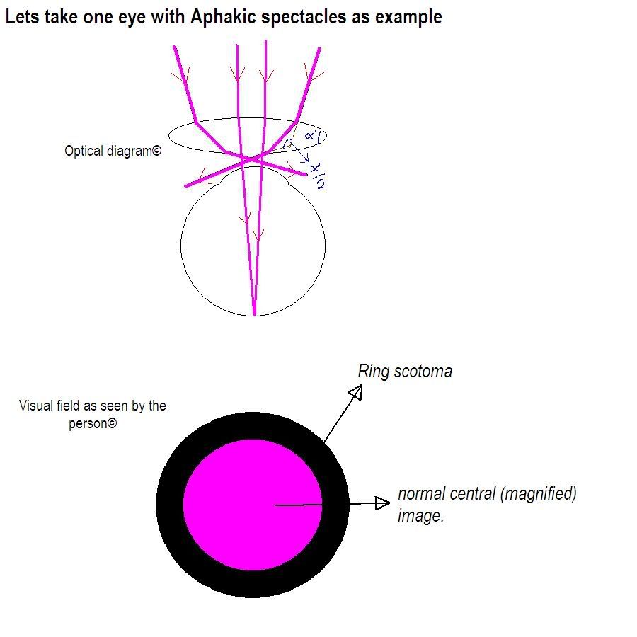
  2. Roving Ring Scotoma: The edge of a convex lens acts as a prism and the higher the power of the convex lens the greater is the prism angle (alpha). The light falling on the prism bends towards its base by an angle alpha/2 , therefore, greater the angle alpha the more will be the bending. In aphakic spectacles, the angle alpha being large, the light falling at the edge of the lens bends towards the center of the lens (base of prism) and does not reach the pupil and is, therefore, not seen. This results in an area of the visual field which is not visible to the patient, or scotoma. And because the edge of the lens is present all around the lens like a ring, so it gives rise to a ring shaped scotoma. The position of this scotoma is not fixed in the visual field because the eye keeps moving (or roving) in relation to the aphakic spectacle. Hence, the result is a roving ring scotoma. roving ring scotoma
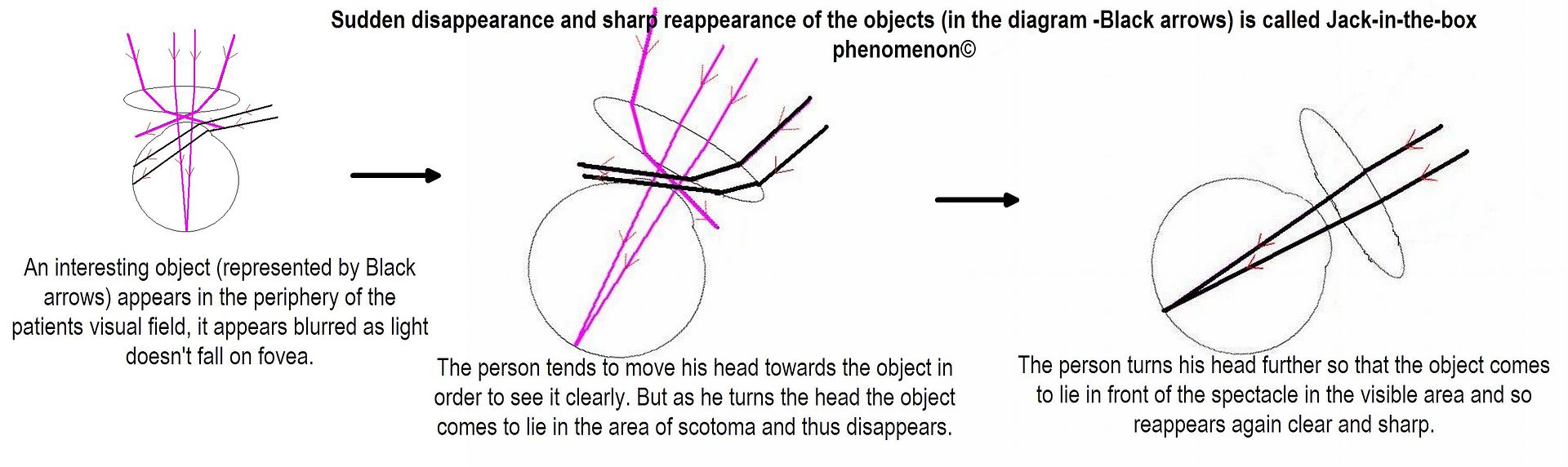
  3. Jack-in-the-box Phenomenon: The presence of the above scotoma leads to another interesting phenomenon. If an interesting object appears in the periphery of the patients visual field, it appears blurred (because the light is passing from the side of the spectacle frame). The person tends to move his head towards the object in order to see it clearly. But as he turns the head the object comes to lie in the area of scotoma and thus disappears. As he turns his head further so that the object comes to lie in front of the spectacle in the visible area and so reappears again clear and sharp. This sudden disappearance and sharp reappearance of the objects is called jack-in-the-box phenomenon. jack in box phenomenon
  4. Pin Cushion Effect: The magnification of image is more at the periphery of the lens due to prism effect. Therefore, all the objects appear stretched out at the corners like a pin-cushion. Pin_Cushion
  5. Spherical Aberrations: The light converges more near the edge of the lens than at the center so the rays of light falling near the edge are brought to focus in front of the rays falling at the center. This results poor quality of image despite appropriate correction of refractive error.
  6. Chromatic Aberrations: The shorter the wave-length the more is the refraction a ray of light undergoes. Therefore, in VIBGYOR the violet end undergoes greater refraction than the red end of the spectrum. This causes diffraction of light and makes the edges of white object appear rain-bow colored.

Saturday, February 21, 2009

Cataracts-Few interesting types

Any opacity of the lens or loss of transparency of the lens that causes diminution or impairment of vision is called Cataract.

christmas tree cataract

  • Oil Drop Cataract  -In Galactosemia.Accumulation of dulcitol (galactose reduction product) within the lens cells leads to increased intracellular osmotic pressure and fluid influx into the lens.Early cataracts reversible with dietary restriction of galactose. Zonular or nuclear opacity with increased refractive power of the nuclear portion causes an "oil droplet" appearance on retroillumination. 

oil droplet cataract

  • Cerulean (blue-dot) cataract -Down's syndrome.

blue dot cataract

  • Rosette Cataract -Traumatic origin.

traumatic cataract

  • Snow Storm or Snow Flake Cataract -In Diabetes . 

snowflake cataract 

  • Sunflower Cataract -In Wilson’s Disease .

snow flower cataract

  • Polychromatic luster-bread crumb appearance -Complicated cataract .

polychromatic lustre 

  • Zonular cataract or Lamellar cataract  (Cataract with riders) -the most common type of Congenital cataract .

lamellar cataract

  • Morgagnian cataract - one type of Hypermature senile cataract . The cortex has turned into milky liquid and the nucleus is displaced inferiorly. 

morganiancataract 

  • Glass Blower’s Cataract -Due to Infrared irradiation.

Tuesday, September 23, 2008

Congenital cataract -causes

Causes of Congenital Cataract

  1. Prenatal (intra-uterine) infection e.g. rubella, cytomegalovirus, syphilis.
  2. Prenatal (intra-uterine) drug exposure e.g. corticosteroids, vitamin A.
  3. Prenatal (intra-uterine) ionizing radiation e.g. x-rays.
  4. Prenatal / peri-natal metabolic disorder e.g. maternal diabetes.
  5. Hereditary (isolated - without associated eye or systemic disorder) e.g. autosomal dominant inheritance.
  6. Hereditary with associated systemic disorder or multi-system syndrome.
    a) Chromosomal e.g. Down’s syndrome (trisomy 21), Turner’s syndrome.
    b) With skeletal disease or muscle disorder e.g. Stickler syndrome, Myotonic dystrophy.
    c) With central nervous system disorder e.g. Norrie’s disease.
    d) With renal disease e.g. Lowe’s syndrome, Alport’s syndrome.
    e) With mandibulo-facial disorder e.g. Nance-Horan cataract-dental syndrome.
    f) With dermatological disorder e.g. Congenital icthyosis, Incontinentia pigmenti.